21 August 2012

Should Replacement Organs be Made, Grown, or Stolen?

Xenotransplantation and organ engineering offer different solutions to the organ crisis, but they share similarities. After decades of research, both fields are in the middle of important clinical trials involving simpler tissues and organs, but complex ones like lungs or liver remain a distant goal. “I think we’re still 2 decades away from something that’s clinically realizable,” says Niklason. _TheScientist
In advanced countries, people are living longer. Citizens are making greater and greater demands from medical technology -- some people think they should live forever. But the human body eventually malfunctions and wears out, eventually coming to an end in cascading failure. But what if tissues and organs could be replaced as they began to show early signs of trouble -- before they could trigger the inexorable cascade to death?
Today, the organ shortage is an even bigger problem than it was in the 1980s. In the United States alone, more than 114,000 people are on transplant lists, waiting for an act of tragedy or charity. Meanwhile, just 14,000 deceased and living donors give up organs for transplants each year. The supply has stagnated despite well-funded attempts to encourage donations, and demand is growing, especially as the organs of a longer-lived population wear out. _TheScientist
There are not enough human cadaver organs to supply the need. The state of the art in both artificial organs and in lab-grown organs and tissues, is years away from large scale application.

So what about stealing the organs? I am not talking about stealing from humans -- as is done in China and other corrupt nations not bound by the rule of law. I am referring to taking organs from animals to use in humans -- xenotransplantation.

These animal organs will have to be designed and engineered to be compatible with the human body, to avoid rejection. And just as animals supply much of our food and an increasing amount of fuels and pharmaceuticals, they could also supply life-saving organs.
Pigs could provide all the organs that we need. They are the right size, and we already have the infrastructure to breed them in large numbers. For decades, people have been fitted with heart valves from pigs, and diabetics injected themselves with pig insulin before we learned how to synthesize the human version of the hormone. Whole-organ transplants, however, are another matter.

The human immune system does not take kindly to the presence of a pig organ. A ready-made armada of antibodies recognizes a sugar molecule called alpha-1,3-galactose (α-gal), which coats the surface of pig blood vessels but is absent from human tissues. The antibodies activate a squad of proteins that make up the complement system, which punches holes in the membranes of the foreign cells on contact. “When I started in the field around 15 years ago, if you put a pig organ into a primate, it was lost in a matter of minutes,” says David Sachs, an immunologist at Massachusetts General Hospital.

Cooper first discovered the α-gal problem in 1992, but it took him until 2003 to fix it. He and others engineered pigs without the α-1,3-galactosyltransferase gene that produces the α-gal residues. In addition, the pigs carry human cell-membrane proteins such as CD55 and CD46 that prevent the host’s complement system from assembling and attacking the foreign cells. “It took 10 years for those pigs to become available, but they made a big difference,” says Cooper.

...While some scientists struggle to get human bodies to accept pig organs, others are attempting the more ambitious feat of engineering human organs from scratch. Such organs, grown from a patient’s own cells, should avoid the problems of immune rejection that plague the field of xenotransplantation. “Cartilage, skin, and bone are already on the market. Blood vessels are in clinical trials. The progress has been really gratifying,” says Laura Niklason of Yale University.

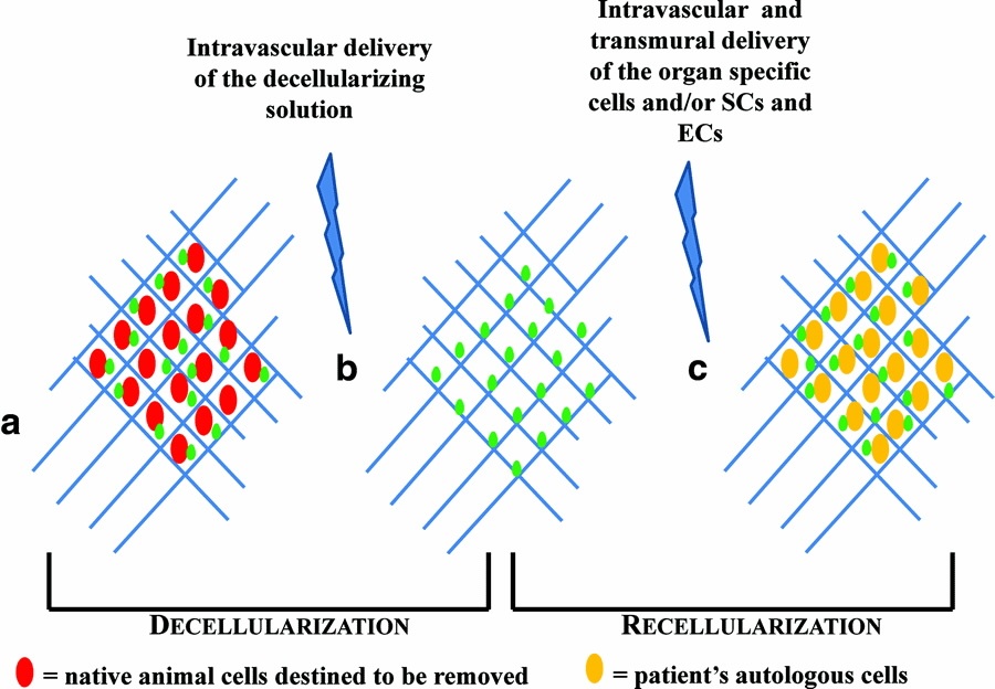
...In 2008, Harald Ott of Massachusetts General Hospital and Doris Taylor of the University of Minnesota dramatically demonstrated the potential of organ engineering by growing a beating heart in the laboratory. As physician-scientists, the two often see patients in dire need of transplantation. They started by using detergents to strip the cells from the hearts of dead rats, leaving behind the extracellular matrix—a white, ghostly, heart-shaped frame of connective proteins like collagen and laminin. Ott and Taylor used this matrix as a scaffold. They seeded it with cells from newborn rats and incubated it in a bioreactor—a vat that provides cells with the right nutrients, and simulates blood flow. After 4 days, the muscles of the newly formed heart began contracting. After 8 days, it started to beat.

...Ott and Taylor’s groundbreaking feat has since been duplicated for several other organs, including livers, lungs, and kidneys. Rodent versions of all have been grown in labs, and some have been successfully transplanted into animals. Recellularized organs have even found their way into human patients. Between 2008 and 2011, Paolo Macchiarini from the Karolinska Institute in Sweden fitted nine people with new tracheas, built from their own cells grown on decellularized scaffolds. Most of these operations were successful (although three of the scaffolds partially collapsed for unknown reasons after implantation). Decellularization has one big drawback: it still depends on having an existing organ, either from a donor or an animal. Frustrated by the wait, Macchiarini tried a different approach. In March 2011, he transplanted the first trachea built on an artificial, synthetic polymer scaffold. His patient, an Eritrean man named Andemariam Teklesenbet Beyene, had advanced tracheal cancer and had been given 6 months to live. “He’s now doing well. He’s employed, and his family have come over from Eritrea. He has no need for immunosuppression and doesn’t take any drugs at all,” says Macchiarini. A few months later, he treated a second patient—an American named Christopher Lyles—in the same way, although Lyles later died for reasons unrelated to the transplantation.

...Whether the scaffold is natural or artificial, clinicians need to seed it with patient’s cells. For bladders or tracheas, it is enough to collect these from a small biopsy. That will not work if the organ is diseased, or if it’s a complex structure of multiple tissue types, or, as in the heart, if its cells are naturally reluctant to divide. In such cases, clinicians will need either stem cells, which can divide and differentiate into any cell type, or progenitor cells that are restricted to specific organs. Since 2006, one source of stem cells has been adult tissues, which scientists can now reprogram back into a stem-cell like state using just a handful of genes. These induced pluripotent stem cells or iPSCs, could then be coaxed to develop into a tissue of choice. “For me, the cells have always been the most difficult part,” says Vacanti, “and I’d say the iPSCs are the ideal solution.” _The Scientist

Labels: ,

Bookmark and Share

28 June 2012

Growing Replacement Organs in the Lab: Wake Forest University's Anthony Atala

This article was originally published on Al Fin Longevity blog

[Anthony] Atala currently heads up more than 300 researchers in the Wake Forest University lab who are working on growing more than 30 different organs and body tissues.

In one trial for the U.S. Armed Forces, his team is collecting healthy skin cells from injured soldiers, processing them, and then spraying them onto battle wounds as a tailored treatment for healing. For deeper wounds, they are in the process of developing an ink jet printer that scans a wound and creates a custom map of the defect.

"After the scan, the printer can go back and print multiple layers of cells right over the wound," Atala said.

The idea of using a patient's own cells rather than relying on those of a donor is important because it eliminates the need to find a "match." For any transplant procedure there is a concern that tissues from a donor will be rejected by a recipient's body. _ABC_via_NBF

There are many challenges to creating lab-grown replacements for human organs. But the promise of being able to create a perfect tissue match replacement organ, and no longer being forced to wait at the bag of the organ donour line, is simply too great a promise to ignore.
By the early 1990s, tissue engineering had become an established field of investigation (30). Concurrently, adult stem cells and ESC were isolated in animals (31, 32) and humans (33), and the advent of nuclear transfer technology made animal cloning possible (7, 34–36). These apparently distinct fields of science had one unifying concept, namely the regeneration of living and functioning body parts destined to replace diseased or damaged cells, tissues, or organs (7). In 1999, the term “regenerative medicine” was coined to describe the use of natural human substances, such as genes, proteins, cells, and biomaterials to regenerate diseased or damaged human tissue (4, 7). It is important to note that the terms tissue engineering and regenerative medicine are not synonymous. The term regenerative medicine is used to define a field in the health sciences that aims to replace or regenerate human cells, tissues, or organs to restore or establish normal function (37). The process of regenerating body parts can occur in vivo or ex vivo and may require cells, natural or artificial scaffolding materials, growth factors, or combinations of all three elements. In contrast, the term tissue engineering is narrower in scope and strictly defined as manufacturing body parts ex vivo, by seeding cells on or into a supporting scaffold. _Excerpted from: Regenerative Medicine and Organ Transplantation: Past, Present, and Future (Atala et al)
Since the early days of tissue engineering, advances in stem cell science, genetic engineering, tissue engineering, 3D printing, and related fields, have given the field of regenerative medicine new powers that were not previously imagined. While all of those sciences continue to advance, it remains for the regenerative medicine specialist to bring them all together and create a new state of the art in tissue and organ transplantation.
One useful "shortcut" in creating new organs from a patient's own tissues, is the use of "acellular" scaffoldings. A donour organ is stripped of its cells, leaving only the supporting acellular matrix scaffolding, including vascular matrix. This scaffolding is then seeded with replacement cells and growth factors for the tissue being replaced, ie vascular, renal, hepatic etc.
A significant advancement in the field of bioscaffold design has been the utilization of decellularized tissue as the three-dimensional scaffold in tissue engineering strategies.11 Our laboratory has previously reported the successful decellularization of porcine aortas and urinary bladder submucosa for use as scaffolds for cell seeding.2, 12 These decellularized aortas were seeded with endothelial progenitor cells and implanted into sheep, and the neovessels remained patent for more than 4 months.2 However, effective decellularization of thicker organs and tissues has been very difficult to achieve due to inefficient penetration of the decellularization solution into the organ. More recently, Ott et al. have developed a more effective method for organ decellularization.13 They have shown that by perfusing a detergent solution through the vascular network rather than relying on agitation and diffusion alone, the entire mouse heart could be decellularized and used as a scaffold for tissue engineering. However, cell seeding of three-dimensional, naturally derived scaffolds presents additional challenges.14 For example, to achieve a recellularized human liver adequate for clinical use, one needs to transfer approximately 10 × 1010 liver cells into the scaffold. So far, such a task has not been successfully achieved. Although perfusion bioreactors have been developed to address cell seeding problems,15, 16 cell seeding across the entire thickness of the scaffold has been limited due to the lack of intrascaffold channels.

The goal of our study was to develop a novel scaffold that human liver cells could readily enter in order to repopulate the scaffold volume. We report the production of such a scaffold via a decellularization process that preserves the macrovascular skeleton of the entire liver while removing the cellular components. The intact vascular tree is accessible through one central inlet, which branches into a capillary-like network and then reunites into one central outlet. Human fetal liver and endothelial cells were perfused through the vasculature and were able to repopulate areas throughout the scaffold by engrafting into their putative natural locations in the liver. These cells displayed typical endothelial, hepatic and biliary epithelial markers, thus creating a liver-like tissue in vitro. This technology may provide important tools for the creation of a fully functional bioengineered liver that can be used as an alternative for donor liver transplantation. _Hepatology 2011 (Atala et al)

Ideally, one would wish to grow and/or print the entire organ in the lab, but the intricate 3D complexity of the intercellular scaffolding of many organs makes such a task very difficult at this time.

Labels:

Bookmark and Share

02 March 2012

Printing Zombies


We already have "printers" capable of printing entire houses. There is the Cornell University printer that prints 3-D flying insect robots. A Drexel University paleontologist is building dinosaur robots out of printed 3-D dinosaur bones.

An elderly woman was a recent surgical recipient of a 3-D printed replacement jawbone. And we are not that far away from printing 3-D replacement tissues and organs using tissue printers.

San Diego startup Organovo is printing muscle tissue with a 3-D printer, aiming to create working muscle.
Unlike some experimental approaches that have used ink-jet printers to deposit cells, Organovo's technology enables cells to interact with each other much the way they do in the body. They are packed tightly together and incubated, prompting them to adhere to each other and trade chemical signals. When they're printed, the cells are kept bunched together in a paste that helps them grow, migrate, and align themselves properly. ­Muscle cells, for example, orient themselves in the same direction to create tissue that can contract.

So far, Organovo has made only small pieces of tissue, but its ultimate goal is to use its 3-D printer to make complete organs for transplants. Because the organs would be printed from a patient's own cells, there would be less danger of rejection. _Technology Review
Organovo will first print human tissues of various types to be used in pharmacological research, to replace animal models and other cruder forms of testing new drugs. They will use the income from sales of these tissue models to drug companies, in order to fund their replacement organ printing research.

But do you see where all of this is leading? First you print the bones, for assembling the skeleton. Then you print the muscles, to allow movement. The organs and blood vessels are then printed and assembled together. The only thing left to add is the brain -- the robotic zombie controller.

While cognitive scientists are almost able to create a zombie brain, they are still decades away from creating a realistic human brain. You should not be discouraged by this, since as long as we remove the cannibalistic instincts of our printed zombies, and train them to be docile and obedient, they can be used for many constructive purposes.

Consider the advantages of being able to use the car-pooling lane in your daily commutes. Or just imagine the surprised looks on your friends' faces when you show up at a party in the company of 2 or 3 zombies who look and are dressed exactly like you!

Just think of the many uses to which trial lawyers could put these zombies in class action lawsuits! Imagine the sympathy they could induce in naive jurors who didn't know any better. Why, one might even be elected president of the USA! It would not be unprecedented.

Anyway, give it some thought. We may not be that far away from such a brave new world, and you want to be prepared.

First published at Al Fin Potpourri

Labels: , ,

Bookmark and Share

22 February 2012

Advances in Repairing Infarcted Myocardial Tissue


UCSD scientists have developed an injectable gel to reinvigorate damaged heart tissue, after a heart attack. Heart failure subsequent to myocardial infarction is a significant cause of death in adults of advanced nations. Tissue replacement and revitalisation treatments such as this, promise to add healthy years to the lives of tens of millions of people, worldwide.
Researchers claim to have developed a new injectable hydrogel which they say could be used to repair tissue damaged by heart attacks.

A team at the University of California, led by Karen Christman, hopes to bring the gel to clinical trials within the next year, the latest edition of Journal of the American College of Cardiology reported.

Therapies like the hydrogel would be a welcome development, Christman explained, since there are an estimated 785,000 new heart attack cases in the US each year, with no established treatment for repairing the resulting damage to cardiac tissue.

The hydrogel is made from cardiac connective tissue that is stripped of heart muscle cells through a cleansing process, freeze-dried and milled into powder form, and then liquefied into a fluid that can be easily injected into the heart.

Once it hits body temperature, the liquid turns into a semi-solid, porous gel that encourages cells to repopulate areas of damaged cardiac tissue and to preserve heart function, according to Christman.

The hydrogel forms a scaffold to repair the tissue and possibly provides biochemical signals that prevent further deterioration in the surrounding tissues. _HinduBusinessLine
Study abstract
Abstract of earlier study

This type of cellular replacement and tissue replacement is important if we are to be able to stretch out the useful lifetimes of genetically flawed bodies. Prevention of disease is better than treatment. Genetic re-design to reduce vulnerabilities may be best of all. Stay tuned.

Labels:

Bookmark and Share

08 November 2011

A Step Toward Curing Parkinson's Disease and New Hope for Curing Many Other Degenerative Diseases of Ageing

Guardian

A research team at Sloan-Kettering have devised a range of methods for converting stem cells into specific differentiated cells. Their most recent triumph was the successful creation of dopamine producing cells from the substantia nigra -- the main part of the brain that degenerates in Parkinson's Disease.
Dr Studer and his colleagues, whose work is published in the journal Nature, found the specific chemical signals required to nudge stem cells into the right kind of dopamine-producing brain cells.

In a series of experiments, the team gave animals six injections of more than a million cells each, to parts of the brain affected by Parkinson's. The neurons survived, formed new connections and restored lost movement in mouse, rat and monkey models of the disease, with no sign of tumour development. The improvement in monkeys was crucial, as the rodent brains required fewer working neurons to overcome their symptoms. _Guardian

The finding brings researchers a step closer to testing a stem-cell-derived therapy in patients with this disorder. "We finally have a cell that seems to survive and function and a cell source that we can easily scale up," says Lorenz Studer, a researcher at the Sloan Kettering Institute and senior author on the new study. "That makes us optimistic that this could potentially be used in patients in the future."

The research also highlights the challenges of generating cells for tissue-replacement therapy, showing that subtle differences in the way the cells are made can have a huge impact on how well they work once implanted.

...While stem-cell researchers had previously been able to create dopamine-producing neurons from human stem cells, these cells did little to alleviate movement problems in animals engineered to mimic the symptoms of Parkinson's. In 2009, Studer and others developed a method of making the cells that more closely mimics the way they form during development. The resulting cells also carry more of the molecular markers that characterize dopamine-producing cells in the brain.

In the new research, published Sunday in the journal Nature, Studer's team found a way to make these cells even more efficiently. This is significant in terms of ultimately testing the therapy in humans; many methods for making specific types of cells are complex and yield small amounts of the desired product. _MIT TechnologyReview
This is a preliminary triumph for regenerative medicine, with every reason to expect that this development can move expeditiously from the animal lab into clinical research. The Sloan-Kettering team has made many improvements in the safe conversion of embryonic stem cells to mature differentiated cells, and have reduced the risk of tumour formation from these cells, when transplanted.

The ultimate goal is to be able to take donour cells from the patient himself, and turn those cells into young and vigorous cells of any cell type that is needed, in as large a number as needed -- even to the point of growing replacement organs from the person's own cells. Researchers are making progress in that area, but the pressing need to treat the growing number of ageing individuals with degenerative conditions may call for stop-gap measures such as the use of embryonic stem cell treatments described above.

Brian Wang has also looked at this story

Labels: ,

Bookmark and Share

07 November 2011

Rejuvenating 100 Year Old Cells: Steps to Regenerative Medicine


"Signs of aging were erased and the iPSCs obtained can produce functional cells, of any type, with an increased proliferation capacity and longevity," explains Jean-Marc Lemaitre who directs the Inserm AVENIR team....The age of cells is definitely not a reprogramming barrier. _SD
Cell Rejuvenation via IPSC

Scientists at the Functional Genomics Institute have taken cells donated by persons older than 100 years, and reprogrammed these senescent cells into pluripotent stem cells and embryonic stem cells. These stem cells can then be differentiated into specialised cells for cell, tissue, and organ replacement therapy -- once the details are worked out.
The researchers have successfully rejuvenated cells from elderly donors, some over 100 years old, thus demonstrating the reversibility of the cellular aging process.


To achieve this, they used an adapted strategy that consisted of reprogramming cells using a specific "cocktail" of six genetic factors, while erasing signs of aging. The researchers proved that the iPSC cells thus obtained then had the capacity to reform all types of human cells. They have the physiological characteristics of "young" cells, both from the perspective of their proliferative capacity and their cellular metabolisms.


Researchers first multiplied skin cells (fibroblasts) from a 74 year-old donor to obtain the senescence characterized by the end of cellular proliferation. They then completed the in vitro reprogramming of the cells. In this study, Jean-Marc Lemaitre and his team firstly confirmed that this was not possible using the batch of four genetic factors (OCT4, SOX2, C MYC and KLF4) traditionally used. They then added two additional factors (NANOG and LIN28) that made it possible to overcome this barrier.


Using this new "cocktail" of six factors, the senescent cells, programmed into functional iPSC cells, re-acquired the characteristics of embryonic pluripotent stem cells.
In particular, they recovered their capacity for self-renewal and their former differentiation potential, and do not preserve any traces of previous aging. To check the "rejuvenated" characteristics of these cells, the researchers tested the reverse process. The rejuvenated iPSC cells were again differentiated to adult cells and compared to the original old cells, as well as to those obtained using human embryonic pluripotetent stem cells (hESC).


...The results obtained led the research team to test the cocktail on even older cells taken from donors of 92, 94 and 96, and even up to 101 years old. "Our strategy worked on cells taken from donors in their 100s. The age of cells is definitely not a reprogramming barrier." He concluded. "This research paves the way for the therapeutic use of iPS, insofar as an ideal source of adult cells is provided, which are tolerated by the immune system and can repair organs or tissues in elderly patients." adds the researcher.


...Inserm's AVENIR "Genomic plasticity and aging" team, directed by Jean-Marc Lemaitre, Inserm researcher at the Functional Genomics Institute (Inserm/CNRS/Université de Montpellier 1 and 2) performed the research. The results were published in Genes & Development on November 1, 2011 _SD
The first use of this new regenerative technology is likely to be cell replacement therapy. But as the methods for growing replacement tissues and organs in the lab are perfected, the methods should be suitable for producing cells to use in growing replacement tissues and organs for purposes of disease treatment and for treating senescence.

Cross-posted to Al Fin Longevity

Labels: , , ,

Bookmark and Share

09 July 2011

Biosingularity Revolution Pushes Against Economic Collapse

Image via NextBigFuture

A revolution in regenerative medicine, genetic recombineering, synthetic biology, systems biology, and several other points of the biosingularity are pushing ahead despite three years of global economic downturn and counting, since the fall of 2008.

Brian Wang introduces us to the "accelerated evolution machine," pictured above.
Say hello to the evolution machine. It can achieve in days what takes genetic engineers years. So far it is just a prototype, but if its proponents are to be believed, future versions could revolutionise biology, allowing us to evolve new organisms or rewrite whole genomes with ease. It might even transform humanity itself.

...Because biological systems are so complex, it is a huge advantage to be able to tweak lots of genes simultaneously, rather than one at a time, she says. "In almost every case you'll get a different solution that's a better solution."

...By automating selection and using a few tricks, though, it should be practical to screen for far more subtle characteristics. For instance, biosensors that light up when a particular substance is produced could be built into the starting strain. "The power going forward will have to do with clever selections and screens," says Church.

As revolutionary as this approach is, Church thinks MAGE's most far-reaching potential lies elsewhere. He reckons it will be possible to use the evolution machine to make many thousands of specific changes to a cell's DNA: essentially, to rewrite genomes.

At the moment, making extensive changes to even the smallest genome is extremely costly and laborious. Last year, the biologist and entrepreneur Craig Venter announced that his team had replaced a bacterium's genome with a custom-written one (Science, vol 329, p 52). His team synthesised small pieces of DNA with a specific sequence, and then joined them together to create an entire genome. It was an awesome achievement, but it took 400 person-years of labour and cost around $40 million.

MAGE can do the same job far more cheaply and efficiently by rewriting existing genomes, Church thinks. The idea is that instead of putting DNA strands into the machine with a range of different mutations, you add only DNA with the specific changes you want. Even if you are trying to change hundreds or thousands of genes at once, after a few cycles in the machine, a good proportion of the cells should have all the desired changes. This can be checked by sequencing.

...As the technology improves and becomes routine, says Church, it could also be used to alter the cells used for cell-based therapies. Tissue-engineered livers grown from stem cells, say, could have their genetic code altered so that they would be immune to liver-destroying viruses such as hepatitis C. _NewScientist_via_NBF

The "evolution machine" has its work cut out for it, but it may very well speed up some projects which do not depend upon significant transformations of the genome. More sizeable genomic transforms are not likely to be possible using such a simplist approach. But future generations of such machines are likely to grow sophisticated enough to make the work of future Craig Venters much faster and simpler.
Researchers at the LA Children's Hospital built a fully functioning artificial small intestine in mice.

A man from Eritrea was recently given an artificial trachea transplant in Sweden. The trachea was grown on a scaffold inside a bioreactor.

Scientists have isolated the human blood cell progenitor stem cell, which is capable of growing all the various cellular components of the blood system.

Johns Hopkins researchers have identified a "super neural precursor stem cell" which can not only differentiate into specialised brain cells, but can also reproduce itself!

A partial list of companies involved in regenerative medicine research and development

The US has been the world's driver of scientific and biomedical R&D for several decades now. There has been some question as to how long American research could maintain its drive, if the nation's economy was dragged down by dysfunctional governmental economic and regulatory policies. Yet, despite the current US government's apparent war against the private sector, some areas of private R&D are still thriving -- although not as well as prior to the fall of 2008.

It is vital that private sector financing be central to advanced R&D, to prevent the type of politicisation of science which has frozen climatology in an infantile state of biased activism (via GWPF), rather than dispassionate observation and honest hypothesis testing.
Federal domination of science funding has two quite intended consequences: both individual scientists and major universities have become wards of Washington. For decades, academic sociologists have noted that almost all faculty party affiliations are with the Democrats. This is no conspiracy–it is merely like-minded individuals hiring other like minds and voting their best interest. _Forbes

Labels: , ,

Bookmark and Share

14 December 2010

News from the Biosingularity

SD
The drug lenalidomide -- related to thalidomide -- may prove to be one of the first in a long line of revolutionary anti-aging medications to slip through the back door of conventional medicine.
In this study, the team tested the drug in healthy seniors, each of whom were matched in race, gender and national origin to a healthy young adult participant. They found that extremely low levels of lenalidomide - 0.1 μM - optimally stimulated IL-2 production in the young people (21-40 years) roughly sevenfold, but stimulated IL-2 production in patients over age 65 by 120-fold, restoring them to youthful levels for up to five days. At that dosage, the drug also increased IFN-gamma up to six fold in the elderly patients, without suppressing IL-17 generation.

The researchers also found that lenalidomide had many other beneficial effects on the elderly participants' T cells, including better migration throughout the body, more efficient patrolling activity and longer survival after defending the body against an infection. _SD
Not for pregnant women, certainly, but not many women over the age of 65 are getting pregnant accidentally.

Another approach to prolonged youthfulness may come by way of "cord blood", blood from the umbilical cord which is obtained at birth. Apparently cells present in cord blood are capable of producing regenerative factors capable of rejuvenating the aging brain.
Laboratory culture (in vitro) studies examining the activity of human umbilical cord blood cells (HUCB) on experimental models of central nervous system aging, injury and disease, have shown that HUCBs provide a ‘trophic effect’ (nutritional effect) that enhances survival and maturation of hippocampal neurons harvested from both young and old laboratory animals.

“As we age, cognitive function tends to decline,” said Alison E. Willing, PhD, a professor in the University of South Florida’s (USF) Department of Neurosurgery and Brain repair and lead author for a study published in the current issue of Aging and Disease (www.aginganddisease.org) . “Changes in cognitive function are accompanied by changes in the hippocampus, an area of the brain where long term memory, as well as other functions, are located, an area of the brain among those first to suffer the effects of diseases such as Alzheimer’s disease.” _SB
The type of rejuvenation of hippocampal cells demonstrated by the USF researchers suggests that it may be worth our while to keep a culture of HUCBs (or useful substitutes) safe and handy for the sake of our older selves.

Another useful approach to rejuvenating the brain besides pharmaceuticals or stem cell therapies may involve the electromagnetic stimulation of the brain.
Shooting steady pulses of electricity through slender electrodes into a brain area that controls complex behaviors has proven to be effective against several therapeutically stubborn neurological and neuropsychiatric disorders. Now, a new study has found that this technique, called deep brain stimulation (DBS), targets the same class of neuronal cells that are known to respond to physical exercise and drugs such as Prozac.

The study, led by Associate Professor Grigori Enikolopov, Ph.D., of Cold Spring Harbor Laboratory (CSHL), is the cover story in the January 1st issue of The Journal of Comparative Neurology, which is currently available online.

The targeted neuronal cells, which increase in number in response to DBS, are a type of precursor cell that ultimately matures into adult neurons in the brain’s hippocampus, the control center for spatial and long-term memory, emotion, behavior and other functions that go awry in diseases such as Alzheimer’s, Parkinson’s, epilepsy and depression. DBS has been successful in treating some cases of Parkinson’s. And recently, it has also proven to work against other brain disorders such as epilepsy and severe depression. _SB
It is likely that routine maintenance for aging humans in the near future will include a wide range of therapies, including stem cells, EM therapies, rejuvenating drug therapies, and nanotech approaches.

Researchers at Salk and Princeton have discovered new ways in which the oncogene protein p53 is useful in the control of cancer. It seems that besides suppressing the early stages of cancer transformation, p53 also suppresses later stage local spread and distant metastasis.
A close collaboration between researchers at the Salk Institute for Biological Studies and the Institute for Advanced Study found that the tumor suppressor p53, long thought of as the "Guardian of the Genome," may do more than thwart cancer-causing mutations. It may also prevent established cancer cells from sliding toward a more aggressive, stem-like state by serving as a "Guardian against Genome Reprogramming." _PO
This opens the door to the use of p53 enhancing therapies in persons whose cancers are already well established -- in the hope of slowing the progression of the disease to allow other therapies to eradicate it.

Another good source of news on the biosingularity front (as well as other science and technology news) is Nextbigfuture:

New artificial bone material to healing from bony injury

New advanced tools for genetic manipulation and engineering

Pluripotent stem cells from adult tissues finding wider use in research

Anyone who has studied Nanomedicine or followed SENS understands that the tools discussed above are just scratches in the surface of the coming biosingularity. But even such scratches may well mean the difference between life and death, vitality and atrophy, mental sharpness and senility, to some who are reading this.

Labels: ,

Bookmark and Share

22 October 2010

Keeping Abreast (or two) of Regenerative Medicine

Regenerative medicine is based upon thebody's ability to build itself -- and to often re-build itself after injury.  We are seeing breakthroughs in stem cell technologies virtually every week.  New technologies that allow physicians to use a patient's own cells to re-build a lost or damaged body part will avoid problems with immune rejection and ethical objections.  And if the touchstone for the explosion of regenerative medicine happens to be the human breast -- who can complain?

How to Build a New Breast


Cytori’s process for reconstructing or augmenting breasts relies on the recent discovery that human fat contains an amazing concentration of stem cells—cells that can be separated out using a centrifuge. That’s the science part. The artistry comes in when the surgeon makes tiny incisions for depositing the enriched fat cells, building a breast one dot-sized injection at a time like a 3-D pointillist. Here’s how it works.

Step 1 Liposuction

Breast reconstruction usually starts in the abdomen, using liposuction to harvest fat cells. Each liposuction syringe holds about 60 cc (2 fluid ounces) of fat cells and takes five minutes to fill. Repairing the divot caused by an average lumpectomy requires eight to 10 syringes to get about 360 cc of fat tissue. Half the fat is used to create the volume needed to fill the divot and half is processed to isolate stem and regenerative cells. A typical augmentation requires 800 cc (27 ounces) of liposuctioned fat: Volume varies, but in one study 160 cc of injected stem-cell-enriched tissue boosted breast circumference an average of 4 centimeters (1.6 cup sizes).

Step 2 Centrifugation

The liposuctioned fat is injected into the Celution System. ›› The fat cells are then “washed” with proprietary enzymes that break down the scaffolding that holds the fat cells together. ›› Next, a centrifuge separates the fat cells from the stem and regenerative cells, concentrating them into a pellet, which is then extracted. ›› The pellet of cells is added back to some of the liposuctioned fat cells, producing a liquid suspension enriched with stem and regenerative cells and ready for injection.

Step 3 Injection


Using a tool called the Celbrush, the surgeon repeatedly deposits the enriched cells in the breast, either at the site of a lumpectomy or throughout the breast for augmentation or repair of a mastectomy.
With reconstruction patients, the tip on the brush makes tiny cuts that perforate scarred areas, transforming the bed of damaged tissue into a biological mesh. The Celbrush releases 0.5 cc of cell-enriched tissue each time the surgeon moves its control wheel. The process typically takes a couple of hours, depending on the extent of treatment. The deposited tissue bonds quickly to the existing tissue. Within 48 hours, new capillaries and blood vessels entwine through the new cells, supplying oxygen and nutrients to the now-stable tissue. ›› The injection area isn’t painful afterward; patients go home the same day.
Source

More from Brian Wang

Labels: ,

Bookmark and Share

23 August 2010

Advances in Regenerative Medicine

The promise of regenerative medicine involves a future where replacement organs and tissues can be re-grown in a lab from a person's own cells, then transplanted into the person as brand-new, fully functioning replacement tissue. Replacement lung tissue, replacement heart tissue, replacement ligaments and tendons, replacement skin, kidneys, muscle, intestine, bladders, and on and on. But first, scientists have to find a good way to grow millions and billions of healthy stem cells from a person's own cells, and keep them alive long enough to turn them into the proper tissue, and grow them on a proper scaffold into the proper replacement organs.
Investigators from the Massachusetts Institute of Technology (MIT) recently developed a new type of support structure for stem cells, which allows them to remain alive for weeks without using any foreign genetic material.

Generally, substrates for growing stem cells contain animal cells or tissue, but these can easily contaminate the samples themselves, which means that they can lose some of their capabilities.

This is an especially serious consequence for induced pluripotent stem cells, which are biological units that can transform into any type of tissue in the human body.

Only environmental conditions dictate whether they will turn into nerve cells, or into pancreatic tissue.

Due to this amazing differentiation ability they have, these cells hold great promise for treating a number of disorders, such as for example Parkinson's, multiple sclerosis and spinal cord injuries.

But, in order to make the best of them, researchers need to be able to grow them in sufficiently large quantities, and this is proving to be extremely difficult due to the lack of proper substrates.

One of the main issues in this field of research is the fact that human stem cells are now grown with the aid of cells or proteins derived from mice embryos. If these foreign chemicals would interact with the human body, they would definitely cause an allergic reaction.

Thanks to the MIT collaborations, which includes biologists, materials engineers and chemists, scientists now have a synthetic surface that features no material from mice or other animals.

The data the team recorded of the new surfaces show that they promote and sustain “all-natural” stem cell growth and reproduction for at least three months. Longer periods are also possible, the group says.

Another impressive feat the MIT experts achieved with their new material is the fact that it allows for researchers to separate colonies of identical cells from each other. The surface allows single cells to form colonies of cells of that type with considerable ease.

Details of the new investigation appear in the August 22 issue of the esteemed scientific publication Nature Materials, e! Science News reports. _Softpedia
Another report from ScienceDaily

“For therapeutics, you need millions and millions of cells. If we can make it easier for the cells to divide and grow, that will really help to get the number of cells you need to do all of the disease studies that people are excited about,' says MIT postdoctoral associate Krishanu Saha, one of the co-first authors of the paper. 

The work was led by MIT professors Robert Langer, Rudolf Jaenisch and Daniel G. Anderson, in collaboration with Saha and postdoctoral researcher Ying Mei. _Softpedia

This report from Brian Wang on Swiss stem cell research, suggests that mature tissue-derived stem cells can be programmed across germ layer boundaries. This finding hints at the possibility of creating virtually any type of cell or tissue from any other type of tissue -- including easily sampled tissues such as skin or blood.

More: Australian researchers at UNSW have developed a process of inducing pluripotent stem cells, iPS, without the use of viruses or "genetic manipulation". Their aim is to generate brain cells to study and treat degenerative brain diseases.

Cross-posted to Al Fin Longevity

Labels:

Bookmark and Share

02 February 2010

Neuronal Pedophilia for Neurodegenerative Conditions

The old seem to imbibe an elixir of life by associating with younger ones of their kind. Swedish researchers have discovered that transplanted stem cells can rejuvenate degenerative neurons, litterally injecting new life into them.
The new report, co-authored by several international research groups and led by Karolinska Institutet, shows that stem cells transplanted into damaged or threatened nerve tissue quickly establish direct channels, called gap junctions, to the nerve cells. Stem cells actively bring diseased neurons back from the brink via cross-talk through gap junctions, the connections between cells that allow molecular signals to pass back and forth. The study found that the nerve cells were prevented from dying only when these gap junctions were formed. The results were obtained from mice and human stem cells in cultivated brain tissue, and from a series of rodent models for human neurodegenerative diseases and acute brain injuries.

"Many different molecules can be transported through gap junctions," says Eric Herlenius, who led the study. "This means that a new door to the possible future treatment of neuronal damage has been opened, both figuratively and literally."

The international team of scientist, beside Karolinska Institutet, included researchers from Sanford-Burnham Medical Research Institute, Harvard Medical School and Université Libre de Bruxelles.

Their report is published in the Proceedings of the National Academy of Sciences _SD

Stem cells may derive either from embryos or from more mature cells. With improvement in techniques for switching on and off expression of various genes will come the ability to create specific stem cells and tissues from a wide variety of mature cells originating within the patient's own body.

Growth factors produced by stem cells tend to regenerate older cells. Studies involving the use of growth hormones and growth factors directly to rejuvenate aging tissues and organisms tend to support the thesis. Using stem cells as "growth factor factories" obviates the need for repeated injections of hormone or factor. Here is a way to make "old stem cells" work like young stem cells again.

It is an analogous phenomenon to older men or women seeking out younger women (or men) for amorous association -- younger women (or men) disseminate pheromones into the local environment which tend to excite various passions -- including, but not limited to, the sexual ones.

When Gandhi routinely slept (chastely) with two underage girls, no doubt he experienced the benefits of "vital transference" at the same time he was strengthening his ability to resist temptation.

Labels: , ,

Bookmark and Share

09 March 2009

Rebuilding a Damaged Brain and More

After strokes and other types of brain damage, entire areas of brain can die and be replaced by fluid filled cavities or depressions. Scientists at Kings College London are experimenting with a biodegradable polymer matrix that may someday re-build the damaged brain after strokes, abscesses, and other types of brain damage.
Scientists say that the key to the advance, published today in the journal Biomaterials, is the use of a biodegradable polymer called PLGA, which ensures that the stem cells remain in the area of stroke damage and establish connections with surrounding brain tissue. By reducing the number of stray stem cells, the system is likely to be safer as well as more effective than other methods, the researchers add.

...The researchers injected particles of the PLGA polymer loaded with neural stem cells directly into the stroke cavities. Once inside the brain, the particles link up to form complex scaffolds. Modo's team used MRI scans to pinpoint where the stem-cell injections were needed and to monitor the development of new brain tissue. "Over a few days we can see cells migrating along the scaffold particles and forming a primitive brain tissue that interacts with the host brain," says Modo. "Gradually, the particles biodegrade, leaving more gaps and conduits for tissue, fibers, and blood vessels to move into." The next step, he says, will be to add the growth factor VEGF, which should encourage blood vessels to enter the new tissue and speed its development into mature tissue.

...The key to the advance was the ability of the new polymer to encourage the growth and differentiation of the neural stem cells at three different scales, says Modo's colleague Kevin Shakesheff, a tissue engineer at Nottingham University. "At the large scale, it enables the void formed by the injury to get new blood vessels very quickly, which is vital if the new tissue is to survive. At the cellular level, the scaffold surface allows stem-cell receptors to attach to it. And at the molecular level, it will allow cells to mix with the right growth factors." _TechnologyReview
Another fascinating area of brain research involves the use of ultra-short electrical pulses to affect nanopores in the nuclear membranes of neurons, while leaving the nanopores of the cell membrane intact. This specificity appears to have interesting effects on the behaviour of neurons individually and as a group.
When an electric field is applied to a cell, a charge starts to build up on the cell membranes. After a few microseconds, the charge is so high that holes (or "pores") start to form in the cell wall, an effect called electroporation. This allows material (in particular calcium ions) to pass through, affecting the function of the cell. With shorter pulses there is not enough time to affect the cell. But electroporation can affect the structures within the cell such as the nucleus, known as organelles.

"Because the organelles are much smaller than the cell itself... they reach their maximum charge much more quickly," Center founder Karl H. Schoenbach explains in an article. " Ending the pulse after the organelles are charged up, within a few hundred nanoseconds but before large pores appear in the cell’s own membrane, lets you focus the electric field’s effects on the organelles, such as the nucleus, while leaving the cell membrane relatively untouched. That, in turn, lets you do the complex and varied things medical science is interested in, such as killing tumor cells or triggering an immune system response."

So on the one hand ultra-short pulses can be used to selectively destroy cancerous cells. But they can also produce much more effective stunning effects.

A paper from the Center on Neuromuscular disruption with ultrashort electrical pulses compares 450-nanosecond pulses with multi-microsecond Taser pulses and found that the shorter pulses were more effective for suppressing voluntary movement, and used less energy. Another study found that even shorter, 60-nanosecond pulses could stun rats.

But the most significant is a paper which found that it was possible to incapacitate cells for a prolonged period -- "our study provides experimental evidence that even a single 60-ns pulse at 12 kV/cm can cause a profound and long-lasting (minutes) reduction of the cell membrane resistance (Rm), accompanied by the loss of the membrane potential." _Wired
I understand if a readers eyes fog up while reading fine print in italics dealing with scientific topics, particularly late at night when they really should be in bed asleep. But it might be worth one's time to contemplate the implications of research on ultra-short electrical pulses on neurons individually and in aggregate.

I will come back to both of these topics in the future.

Labels: , ,

Bookmark and Share

22 January 2009

Building New Cellular Infrastructure

Tissue scaffolds are the next big thing for implants of the future. Like the scaffolding we see on construction sites, the nano scaffolds are being created by Ko to reconstruct damaged tissue within the human body. Burn victims would benefit from scaffolds used to regenerate new skin. Those with failing heart valves or damaged nerves could count on scaffolds to regenerate these parts from within the patient’s own body. As healing progresses, the scaffold, being constructed from a biodegradable material, is absorbed and metabolized by the body while slowly releasing drugs to aid in the healing process. _CyborgAge
Almost every part of the body presents opportunities for scaffold bio-engineers to experiment. From the heart to the spine to the skin, all parts of the body eventually wear out and need to be replaced or regenerated. Scientists at UC Berkeley are taking an entirely new approach to bio-scaffold development. They are using viruses (bacteriophages) to build a proteinaceous infrastructure that promotes regeneration of nerve tissue.
Some biological engineers are using scaffolds made of polymers to try to mimic the supportive matrix of real tissue. Seung-Wuk Lee, a bioengineer at the University of California, Berkeley, has turned to viruses instead. "Viruses are smart materials," he says. "Once you construct the genome, you can make billions of phages, and they're self-replicating materials." The phage that Lee is working with, called M13, is long and thin like the protein fibers that make up the cellular matrices inside the body.

First, Lee and his colleague Anna Merzlyak genetically engineered M13 to display nerve-friendly proteins on their outer coats. These proteins are known to help nerve cells proliferate, adhere, and extend into long fiberlike shapes. Next, the researchers grew large numbers of the viruses in bacterial-cell hosts and dropped them into a solution containing neural-progenitor cells. These cells are more fully developed than stem cells but are still young and need coaxing to form new tissues. In the solution, the viruses align themselves like a liquid crystal, says Lee. He and Merzlyak used pipettes to inject the solution into agar, a Jell-O-like cell-culture medium, creating long, nerve-like fibers of the virus interspersed with cells. The progenitor cells then multiplied and grew the long branches characteristic of neurons. Lee says that the phage are well suited to making long, fiberlike structures such as nerve tissue but can also be made into more complex structures by varying their concentration or manipulating their position with a magnetic field. _TechnologyReview
Lee is planning to move to research inside live animals next. He is interested to discover how the immune systems of animals will react to viral construction workers hammering, drilling, and welding new infrastructure deep inside the organism.

For regenerative medicine to take that next big step forward, it will need the ability to grow specific infrastructure for every tissue and organ type that will be replaced or regenerated. Then, scientists will need to integrate growth factors and stem cells into the new matrix, and provide optimal nutrient solution. The prognosis for significant progress in this area is extremely favourable.

Labels:

Bookmark and Share

02 October 2008

Saving the Brain and Spinal Cord With Silicon Nanoparticles + PEG + Hydralazine

Researchers at Purdue University have combined silicon nanoparticles with polyethylene glycol (PEG) and the antihypertensive drug hydralazine, seeking a way to preserve brain and spinal cord function after central nervous system injury and trauma.
A team led by Richard Borgens of the School of Veterinary Medicine's Center for Paralysis Research and Welden School of Biomedical Engineering coated silica nanoparticles with a polymer to target and repair injured guinea pig spinal cords. That research is being published in the October edition of the journal Small....The team then used the coated nanoparticles to deliver both the polymer and hydralazine to cells with secondary damage from a naturally produced toxin. That research was published in August by the journal Nanomedicine.

....In the first study, the researchers coated the nanoparticles with PEG to treat guinea pig spinal cord injuries. The treated spinal cord cells showed improved physiological functioning.

In the second study, the researchers added both PEG and hydralazine, an antihypertension drug, to mesoporous silica nanoparticles. These nanoparticles have pores that can hold the drug, which is later delivered to the damaged cells. The hydralazine was added to fight off secondary damage to cells that occurs after the initial injury.

"When cells are injured, they produce natural toxins," Borgens said. "Acrolein is the most poisonous of these toxins. It's an industrial hazard for which hydralazine is an antidote."

Borgens and his team introduced acrolein into cells and then treated the cells with different combinations of hydralazine and/or PEG delivered by the mesoporous silica nanoparticles....They found that the treatment restored disrupted cell function caused by acrolein.

The team concluded that the use of nanoparticles to deliver both PEG and hydralazine increased the effectiveness of earlier PEG-only treatment by controlling and concentrating release of the drug and the polymer, producing a dual treatment and prolonging the treatment's duration.

...."All ambulances should have PEG on board," he said. "It can probably save thousands of people from more severe head and spinal damage." _PO
As biomedical science develops better treatments for degenerative and infectious diseases, better means to prevent and treat cancer, and better ways to delay and reverse the aging process, more attention will turn to the treatment of traumatic injury. Brain injury is easier to prevent than to recuperate from. Even so, expect more and more uses of nanoscale treatment modalities combined with regenerative therapies in the natural human attempt to turn back the entropy of trauma, time, and life.

More on the use of PEG in head injury

Labels: ,

Bookmark and Share

25 February 2008

Tissue Scaffold News

Tissue and organ replacement--regenerative medicine--is dependent on new developments in stem cells, growth factors, genetic controls, and tissue scaffolds. One approach to tissue scaffold uses nano-polymers.
David Nisbet from Monash University's Department of Materials Engineering has used existing polymer-based biodegradable fibres, 100 times smaller than a human hair, and re-engineered them to create a unique 3-D scaffold that could potentially allow stem cells to repair damaged nerves in the human body more quickly and effectively.

Mr Nisbet said a combined process of electrospinning and chemical treatment was used to customise the fibre structure, which can then be located within the body.

"The scaffold is injected into the body at the site requiring nerve regeneration. We can embed the stem cells into the scaffold outside the body or once the scaffold is implanted. The nerve cells adhere to the scaffold in the same way ivy grips and weaves through a trellis, forming a bridge in the brain or spinal cord. Over time, the scaffold breaks down and is naturally passed from the body, leaving the newly regenerated nerves intact," Mr Nisbet said.___Source
Certainly polymer based scaffolding can be generated quickly, and modified relatively easily. I will be interested to follow developments from this approach.

A special award was given recently to a Yale researcher involved in tissue scaffold development.
Erin Lavik, an assistant professor of biomedical engineering at Yale, was honored recently by the Connecticut Technology Council as one of their 2008 Women of Innovation....Lavik, who was cited for her academic innovation and leadership, focuses her research on developing new therapeutic approaches for the treatment of spinal cord injury and retinal degeneration.

She begins repair of damaged tissues using biodegradable polymers formed into three-dimensional scaffolds that mimic the structure of the tissue. After chemically modifying the scaffold surfaces, she incorporates growth factors that further create an environment for repair.

By combining neural or retinal stem cells with these environments, she is discovering the cues that promote integration and differentiation of the cells into healthy tissue. In a rodent model of spinal cord injury, the seeded scaffold promoted functional recovery allowing the rats to regain a weight-bearing stride. She also collaborated on an implantable system that can form and stabilize a functional network of fine blood vessels critical for supporting tissues in the body.___Eurekalert
Tissue scaffolds are routinely subjected to a variety of testing, in order the achieve the proper combination of properties of mechanics and permeability.
Deformable scaffolds with specific mechanical properties were made by blending flexible, biodegradable polymers.3,4 Labyrinths of pores with specific shapes and interconnectivity were formed into cube-shaped samples using injection molding and 3D printing.5 These prototypes were then cyclically distorted to varying degrees and in several ways: compressed or twisted, for instance. Micro x-ray imaging followed the movement of a contrast dye through the scaffolds as they were manipulated.___Source
Current scaffold-like products being used in the OR include Apligraf, Alloderm, among a growing list of synthetic tissue graft and scaffold products.

The "inkjet" approach to printing tissue and tissue scaffolds is also an active area of research in regenerative medicine--although not ready for the OR yet. The time is certainly coming, when most human organs and tissues will be replaceable with lab-grown stand-ins. We do not need anything fancy. Just something that works.

Labels: ,

Bookmark and Share

14 January 2008

The Quest for Replacement Body Parts

Since 1967, when Christiaan Barnard successfully performed the first successful human to human heart transplant, it has been painfully obvious that there are not enough human hearts available to meet the demand. Surgeons have tried various approaches to replacing damaged and worn out hearts--including baboon hearts, refrigerator sized machine replacement hearts, and most reacently, the AbioCor totally implantable artificial heart system. While the AbioCor's batteries can be rechared through the skin, they will eventually have to be replaced. Machine hearts are subject to failure of various types, and experience with them is still only short term.

Eventually, hearts will be "printed", along with other replacement organs. Other organs, such as the urinary bladder, have already been synthetically produced and implanted. But the heart's fibrous skeleton is too complex for scientists to mimic in the lab--to this point.

So lab scientists are learning how to scavenge heart skeletons wherever they can. University of Minnesota scientists have taken dead animal hearts and removed the dead cells--leaving the fibrous infrastructure. By injecting immature heart cells into the scaffolding--in a stepwise manner--they were able to revitalize the heart to the point of beating.
The team took a whole heart and removed cells from it. Then, with the resulting architecture, chambers, valves and the blood vessel structure intact, repopulated the structure with new cells.

"We just took nature's own building blocks to build a new organ," says Dr Harald Ott, a co-investigator who now works at Massachusetts General Hospital. "When we saw the first contractions we were speechless."

The work has huge implications: "The idea would be to develop transplantable blood vessels or whole organs that are made from your own cells," said Prof Doris Taylor, director of the Centre for Cardiovascular Repair, Minnesota, principal investigator.

The method could be used to grow liver, kidney, lung and pancreas, indeed virtually any organ with a blood supply.
Telegraph

In the meantime, researchers continue to work on alternatives--including artificial hearts that spin like a turbine, producing a constant blood pressure rather than a pulse. Such turbine hearts are said to be efficient in small sizes, making it easier to fit size constraints. Such hearts would still have the problem of requiring a power supply.

Rather than replacing the heart, methods of regenerating the existing heart are being developed. Techniques of injecting stem cells into the patient's heart have already produced positive results in some cases. Likewise, procedures that attach "sheets of muscle blasts" to the patient's heart have been successful in Japan. Clearly, it would be preferable if the patient's own heart can serve as a scaffold for cell replacement.

In several pathological processes, however, the underlying structure of the patient's heart has been rendered dysfunctional. Without extensive remodeling surgery, the heart's infrastructure has to be replaced.

In approaching heart replacement, patients, physicians, and families have to weigh the benefits and risks. With the rapid growth in viable choices for replacement, this process will necessarily become more detailed and informed.

Bioprinting organs, and other ways of synthetically reproducing human organs, will be very expensive for a long time. That expense will push experimentation in different directions.

Labels: , ,

Bookmark and Share

20 December 2007

Repair That Heart! Clever Muscle Cell Therapy Bypasses Transplant--Regenerative Medicine Comes of Age

Osaka University Hospital researchers have grown sheets of a patient's muscle cells (myoblasts) in the laboratory, then in the operating room applied these myoblast sheets to the patient's failing heart.
...the researchers first took about 10 grams of muscle from one of the patient’s thighs. Myoblast cells (a type of muscle stem cell) were then extracted from the muscle tissue, placed in a culture solution and grown into 50-micron-thick sheets measuring about 5 centimeters (2 inches) in diameter. Several layers of myoblast sheets were then applied to the surface of the impaired heart, where they helped strengthen the muscle and restore cardiac function.

Within months, the patient’s pulse rate and cardiac output (the amount of blood pumped from the heart with each contraction) returned to normal levels. The patient’s ventricular assist device was removed in September, and doctors say he will be able to lead a normal life after being released from the hospital at the end of this month.
Pink Tentacle

Clearly this procedure required extensive preparation and post-surgical rehabilitation. But consider what a heart transplant requires. Besides waiting for years for a donor, the surgery, the rehab, then a lifetime of anti-rejection drugs. No, clearly the myoblast approach is the winner--if it works.

Regenerative medicine is the process of using advanced cellular biology techniques to "refurbish" a failing organ. The heart is an obvious candidate for regenerative medical approaches. The kidneys, lungs, liver, and pancreas are others. Eventually, it will be possible to regrow any organ or tissue from adult stem cells (ASCs) using the appropriate scaffolding and growth factors, and knowing how to turn the proper genes on and off at the right times.

Kudos to the Osaka team.

Labels: ,

Bookmark and Share

05 December 2007

Chimeric Mice Help in Drug Development

Chimeric mice developed with human liver cells growing inside mouse livers will prove to be valuable toxicity screens for pharmacologic development, improve understanding of infectious disease, and promote regenerative medicine.
The work, which will be published in this week's online edition of the Proceedings of the National Academy of Science also holds promise for a better understanding of infectious diseases that affect the liver. "It is basically impossible to grow human hepatocytes in the lab, which was a big hurdle for the study of viruses such as hepatitis A and hepatitis B," says senior author Inder Verma, Ph.D., a professor in the Laboratory of Genetics.

But most importantly, Bissig says, the mice will be an invaluable tool to advance regenerative medicine. "Many inherited disorders affecting liver metabolism could be cured if only five percent of all hepatocytes would express the missing enzyme," he says.
Salk Institute

Hat tip Medgadget

Labels: , ,

Bookmark and Share

09 October 2007

Tissue Engineering of Blood Vessels and Other Tissue

Tissue engineering is beginning to yield some useful products. Using skin tissue, scientists and bio-engineers can grow blood vessels for replacement and bypass surgeries.
From a snippet of a patient’s skin, researchers have grown blood vessels in a laboratory and then implanted them to restore blood flow around the patient’s damaged arteries and veins.

It is the first time blood vessels created entirely from a patient’s own tissues have been used for this purpose, the researchers report in the current issue of The New England Journal of Medicine.

Cytograft Tissue Engineering of Novato, Calif., made the vessels, in a process that takes six to nine months. Because they are derived from patients’ own cells, they eliminate the need for antirejection drugs. And because they are devoid of any synthetic materials or a scaffolding, they avoid complications from inflammatory reactions.
Source
Better scaffolds for growing tissues in the lab are being developed. The gel scaffold pictured above incorporates microchannels for nutrient fluid supply to the growing tissues--an artificial "blood" vessel.
The researchers have engineered tiny channels within a water-based gel that mimic a vascular system at the cellular scale and can supply oxygen, essential nutrients and growth factors to feed individual cells. The so-called gel scaffold can hold tens of millions of living cells per milliliter in a 3-D arrangement, such as in the shape of a knee meniscus, to create a template for tissue to form.

In theory, the system could accommodate many kinds of tissue.

"A significant impediment to building engineered tissues is that you can't feed the core," said Abraham Stroock, Cornell assistant professor of chemical and biomolecular engineering and one of the paper's senior authors. "Simply embedding this mimic of a microvascular system allows you to maintain the core of the tissue during culture." Gel scaffolds, he said, "are the culture flasks of the future."

The embedded microchannels allow fluid with oxygen, sugar and proteins to travel through the system. The researchers can control the distributions of these solutes over both time and space within the developing tissue, allowing the fine-tuning of the biochemical environment of the cells while the tissue develops. For example, the tissue may need to develop into bone on one side and cartilage on the other. Now the researchers can supply the right nutrients and proteins to certain parts of the growing tissue to ensure an intended outcome.
Source

As scientists and bio-engineers learn to mimic normal in vivo tissue growth processes in the lab, we will have more and better tissue and organ replacements available for transplant and regenerative purposes. Eventually, we will be able to grow better tissues and organs than the originals. Tissues more resistant to wear and degradation. Stronger muscles. More efficient nerves that are resistant to degenerative influences. Blood vessels that resist occlusive processes. Bones less prone to breaking etc.

Labels: ,

Bookmark and Share

04 September 2007

Introducing the grobyC--the Inverse of a Cyborg

The problem of devising linear actuators for autonomous robots is an interesting one. Several artificial substitutes for muscle have been devised and tested. Scientists from South Korea have decided to avoid the substitutes and go straight to the real thing--using actual living muscle tissue as robot actuators!

A cyborg is the use of machines and artifacts to replace living tissue in a living organism. A grobyc is merely the inverse of a cyborg--the use of living tissue to replace machinery in a machine. The scientists from South Korea have created a grobyc!
According to Chemical Science, Sukho Park of the Nano/Micro System Laboratory at the Seoul National University and his colleagues "made the robot by growing heart muscle tissue from a rat onto tiny robotic skeletons made from polydimethylsiloxane (PDMS)."

You can see above how the scientists prepared their microrobot: (a) Single heart cells isolated from neonatal rat heart. (b) PDMS structure prepared for culture of cardiomyocytes on its surface. (c) Primary cardiomyocytes on the culture dish containing the PDMS structure. (d) Culture of cardiomyocytes. (e) Transfer of PDMS structure into a new culture dish to observe movement. (f) Schematic image to observe vertical movement. (g) Microscopic image of vertical view. (h) Schematic image to observe lateral movement. (i) Microscopic image of lateral movement. (Credit: Sukho Park and his colleagues)
primidi

Okay, this is not the first grobyc. Back in 2004, scientists used an array of 25,000 rat brain neurons to fly a simulated F-22 fighter jet. The scientists from South Korea have extended the concept to muscle as linear robot actuators. That is quite clever.

Nanotechnologists, if they are smart, borrow shamelessly from biological mechanisms and proofs of concept. It should not surprise us that roboticists would likewise borrow from biology to solve difficult problems in the actuation of autonomous robots.

Muscle is an excellent linear actuator, and is powered by simple nutrients that can be obtained easily. When muscles grow weak or scarred, losing their normal function, a good method of replacing them within living organisms--such as people--would be quite convenient. Fortunately, Pittsburgh scientists have located adult stem cells within the walls of blood vessels that fit the task perfectly. Palliatives for muscular dystrophy or scarred hear walls may be within reach.

Back to the grobyC. How far, do you think, can scientists go in using living tissue and biological ingredients in robots and other machines? The South Korean scientists used cardiomyocytes from the Sprague-Dawley rat. What if they wanted to use the entire heart of the rat as a mechanical pump? Or the digestive system as a way of processing nutrients for the pump and actuators? Or the rat brain as a controller for the grobyc, as the US investigators did in 2004?

At what point does the grobyc become cyborg?

Labels: , , ,

Bookmark and Share
Older Posts
Al Fin Main Page
Enter your Email


Powered by FeedBlitz
Google
WWW AL FIN

Powered by
Blogger

``