14 February 2012

This'll Kill Ya! Affirmative Action Doctors Meet Lower Standards

1. For those students applying to medical school with average GPAs (3.40 to 3.59) and average MCAT scores (27-29), black applicants were almost three times more likely to be admitted than their Asian counterparts (85.9% vs. 30%), and 2.4 times more likely than their white counterparts (85.9% vs. 35.9%). Likewise, Hispanic students with average GPAs and average MCAT scores were about twice as likely to be accepted as white applicants (68.7% vs. 35.9%), and more than twice as likely as Asian applicants (68.7% vs. 30%).

2. For students applying to medical school with slightly below average GPAs of 3.20-3.39 and slightly below average MCAT scores of 24-26 (first column in the table), black applicants were more than 8 times as likely to be admitted as Asians (67.3% vs. 7.7%), and more than 5 times as likely as whites.

...U.S. medical schools must be considering race as one important factor in admissions, at least for preferred minority groups (blacks and Hispanics) over non-preferred minority groups (Asians) and whites?
_Carpe Diem
When you are attended by a physician of any race, you have to right to expect that that physician had to meet the high level of standards in acceptance, training, and certification that any other physician had to meet. But with affirmative action in admissions and hiring at all levels of medical school, residency, and hospital positions, the more likely reality is that minority physicians were not held to the same high standards as other trainees and job applicants.
But more disturbing even than the finding that medical schools seem to be admitting less-qualified students on the basis of race and ethnicity is that many of these students can't pass their licensing exams, despite greater resources directed toward helping them than other students received. At every medical school CEO studied, substantially larger numbers of black students than whites either did not take or failed their initial licensing exams, and, in most instances, failed their subsequent licensing tests as well. _Affirmative Action Doctors Can Kill You
There is a great deal of political pressure to advance unqualified minority candidates in a wide variety of fields, from medicine to law to engineering to police and fire fighting. It is just too bad if you or a member of your family happens to be in a situation where you need the best level of help, but are instead forced to settle for much less.

Here is one example of an affirmative action doctor, whose tragic example only fell into the spotlight by accidental connection to a famous lawsuit (Bakke).
He readily conceded that he would never have been admitted to medical school under the normal standards, but maintained grades of 3.2 to 3.3 on a 4.0 scale.

After residency and earning a master's degree in public health from the University of California at Los Angeles, he moved to Compton and set up his practice in nearby Lynwood.

His professional difficulties began in 1993, at Long Beach Memorial Hospital, when he was accused of mishandling a delivery, and the hospital began monitoring him. He sued, charging racism. In a jury trial, he won $1.1 million in damages, but a judge overturned the verdict.

By 1997, he said he had delivered 10,000 children and performed thousands of abortions. About that time, he added liposuction to his practice.

His personal and professional life then took a further downturn. In 1997, The Associated Press found in court records that he had been sued 21 times for malpractice and had settled some suits with no admission of guilt. He declared bankruptcy and went through the second of two divorces.

In 1997, his license was suspended, for not paying child support, but he continued to practice. The medical board used that as one of more than 90 counts in revoking his license the next year. _NYT
Sure, you could find similar tragic stories from members of virtually any ethnic group, but such things are more likely to happen when the screening standards for a profession are allowed to drop for one reason or another.

There is no way to know whether a physician who is a member of an ethnic minority was accepted under lowered standards, at the level of medical school, residency, or as a professional hire. And it is that uncertainty which should concern anyone who is worried about the quality of care that he or his loved ones are likely to receive, should it be necessary.

In the age of Obamacare -- where more and more employers will be squeezed into dropping their health insurance plans, and where choice of personal physicians is beginning to evaporate away -- the uncertainty is likely to grow acutely worse over time.

Labels: ,

Bookmark and Share

09 June 2009

Magic Helical Bullets to Kill Cancer and Bacteria?

The compound [Fe2L3]4+ is an iron triple helicate with three organic strands wrapped around two iron centres to give a helix which looks cylindrical in shape and neatly fits within the major groove of a DNA helix. It is about the same size as the parts of a protein that recognise and bind with particular sequences of DNA. The high positive charge of the compound enhances its ability to bind to DNA which is negatively charged.

When the iron-helicate binds to the major groove of DNA it coils the DNA so that it is no longer available to bind to anything else and is not able to drive biological or chemical processes. _PO
The interesting structure known as [Fe(2)L(3)](4+) was first researched as a treatment for cancer. But University of Warwick researchers have discovered that the metallomolecule is a particularly potent killer of bacteria -- even bacteria that are resistant to conventional antibiotics.
Initially the researchers focused on the application of this useful property for targeting the DNA of cancer cells as it could bind to, coil up and shut down the cancer cell's DNA either killing the cell or stopping it replicate. However the team quickly realised that it might also be a very clever way of targeting drug-resistant bacteria.

New research at the University of Warwick, led by Dr Adair Richards and Dr Albert Bolhuis, has now found that the [Fe2L3]4+ does indeed have a powerful effect on bacteria. When introduced to two test bacteria Bacillus subtilis and E. coli they found that it quickly bound to the bacteria's DNA and killed virtually every cell within two minutes of being introduced - though the concentration required for this is high. _PO
It will be important for the researchers to learn ways that the compound can be preferentially transported across bacterial membranes, to bacterial DNA. By reducing the necessary concentrations needed to kill the bacteria, and by making the construct preferentially attracted to pathological bacteria rather than to normal human cells, the safety and the efficacy of any possible future treatment using this compound will be enhanced.
The prevalence of antibiotic resistance has resulted in the need for new approaches to be developed to combat previously easily treatable infections. Here we investigated the potential of the synthetic metallomolecules [Fe(2)L(3)](4+) and [Cu(2)(L')(2)](2+) as antibacterial agents.... [Fe(2)L(3)](4+) binds in the major groove and causes DNA coiling... The work described here shows that ... [Fe(2)L(3)](4+) is bactericidal for Bacillus subtilis and Escherichia coli. We demonstrate that [Fe(2)L(3)](4+) binds bacterial DNA in vivo and, strikingly, that it kills B. subtilis cells very rapidly. _IntJnlAntimicrobialAgents
Cytocidal approaches are valuable medically to the extent they can be targeted and controlled -- with only limited damage to normal cells and tissues.

One of the main pillars of the SENS anti-aging approach is the elimination of superfluous and dangerous cells that have outlived their usefulness. The more precisely that one can target cytotoxic molecules, the more useful they will be for anti-aging therapies in addition to more conventional medical disciplines such as oncology and infectious disease.

Labels: , ,

Bookmark and Share

01 June 2009

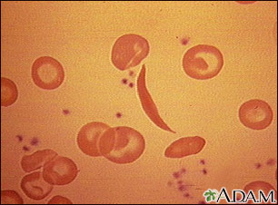
Fast Track Cures for Sickle Cell, Spherocytosis, and Other Blood Diseases

Blood disorders are likely to be the first targets for therapy because corrected cells can easily be transferred back to the patient via bone-marrow transplants. _TechnologyReview
Salk Institute researchers in La Jolla have demonstrated a technique that opens the door to cures for several genetic blood diseases. It involves taking a patient's skin cells, treating them genetically to correct the inherited disorder, next turning the skin cells into induced pluripotent stem cells (iPS), then differentiating those iPS into blood progenitor cells. These progenitor cells can be implanted into the patient to provide a supply of normal blood cells.
"This is an exciting bit of science," says Chris Mason, a professor of regenerative medicine at University College London, who was not directly involved in the research. "It's likely to be the first of a slew of similar papers that may offer hope for conditions where today there is no real therapy, let alone a cure."

So far, Belmonte's approach is applicable only to diseases in which the genetic defect that underlies the disease has been identified. "But there are quite a few of these--and the number will increase," says Mason. Blood disorders are likely to be the first targets for therapy because corrected cells can easily be transferred back to the patient via bone-marrow transplants.

Belmonte adds that in the future, the correction of more-complex genetic disorders might become possible, thereby significantly increasing the number of diseases that might be treated with altered iPS cells. _TechnologyReview
If the bone marrow can be "re-stocked" with viable normal blood progenitor cells, the inherited blood disorder may indeed be cured. The concept can be extended to other inherited and acquired genetic diseases with extensive work.

For parents and health care workers, life-threatening childhood diseases are one of the greatest possible tragedies of existence. Most fatal infectious diseases of childhood have been controlled in the developed world. Now it is time to cure the inherited disorders of childhood that destroy so many lives long before their time.

More from Brian Wang at NextBigFuture on the coming revolution in stem cell replacement therapies.

Labels: ,

Bookmark and Share

02 February 2009

Autologous Stem Cells Hold Most Promise in MS

Medical scientists are learning how to use autologous stem cells -- a person's own stem cells -- to treat serious illnesses such as multiple sclerosis (MS). A recent study at Northwestern University demonstrates an ingenious approach to the use of autologous stem cells:
In clinical trials, a team of scientists led by Richard Burt of Northwestern University in Chicago essentially rebuilt the immune system of 21 adults – 11 women and 10 men – who had failed to respond to standard drug treatments.

First they removed defective white blood cells that, rather than protecting the body, attacks the fatty sheath, called myelin, that protects the nervous system.

The immune systems were then replenished with so-called haemopoeitic stem cells – extracted from the patient's bone marrow – capable of giving rise to any form of mature blood cell.

....After an average follow-up period of three years, 17 of the 21 patients improved by at least one point on a standard disability scale, and none had a final score lower than before the stem cell transplant. _Cosmos
The ability to not only stabilise, but to actually reverse symptoms of advanced MS using autologous cells, is encouraging. Autologous cell therapies are preferred over cell transplants from donors or ESCs, due to the virtual absence of immune system rejection. That is why the new methods of inducing pluripotent stem cells from adult cells such as skin cells, will almost certainly be the preferred method of future medical rejuvenative and regenerative cell and (grown) organ replacement therapies.

Lancet article

Labels: , ,

Bookmark and Share

26 September 2008

A New Age for Stem Cells and Regeneration

A new way of inducing the creation of pluripotent stem cells (iPS) from mature cells is opening the way for a positive deluge of new stem cell research--unrestricted by government funding limitations, access to embryos, or danger of cancerous transformation. Regenerative medicine has never been as promising as it is right now.
Many view the creation of genetically unmodified iPS cells as regenerative medicine's magic bullet. The cells are not derived from embryos, so researchers can circumnavigate the ethical gray areas. And if these cells turn out to be as potent as embryonic stem cells, they could be used to help regrow tissues damaged in conditions ranging from paralysis to Parkinson's disease to diabetes. If they can be grown from a patient's own cells, they could furthermore be transplanted without triggering immune rejection.

...The efficiency, as described in latest edition of the journal Science, is still incredibly low. Out of 1 million adenovirus-infected cells, the researchers ultimately produced just one stable line of stem cells. But the line was genetically unaltered, and when the cells were implanted in mice, they formed a cluster of cells that had differentiated into multiple tissue types (a standard test for pluripotency). When the researchers injected the cells into mouse embryos, the resulting mice had integrated the stem cells into a number of different types of tissue, including tissue in the brain, lungs, and heart. And mice as old as 13 weeks remained tumor-free.

Until now, iPS cells couldn't be compared to embryonic stem cells, since the effects of the integrated virus were unknown. "It was like comparing apples and oranges," Hochedlinger says. Now, however, the potency of the two cell types can be evaluated head to head. "You can really think about doing this in a human setting now, and about making genetically unmodified human cells for modeling or even for therapy."

The finding already has other stem-cell experts thinking about the possibilities. "The paper represents a major breakthrough in reprogramming research and proves to the field that we can reprogram cells directly without viral contamination," says George Daley, a Harvard biologist and stem-cell researcher who was not involved with the research. "It is a major step towards making clinical transplantation of patient-specific cells feasible."

Hochedlinger and his colleagues are now working to increase the efficiency of their adenovirus technique and to repeat their methods to create human iPS cells. "Once we do that," Hochedlinger says, "we can figure out whether [embryonic stem] cells and unmodified iPS cells are really identical to each other or not. I don't know the answer yet." _TechReview
Once the efficiencies of the process are improved, availability of patient-specific pluripotent stem cells should skyrocket, and ethical and technical obstacles to clinical use of stem cells should steadily melt away.

Labels:

Bookmark and Share

13 May 2008

MicroArrays in Research: A Silent Revolution

A Micro-array is a potent tool of biological discovery, first used to study genetic variation, and now rapidly being adapted to a wide range of biochemical research. Briefly, micro-arrays incorporate a large number of biochemical probes onto a single chip, to allow up to thousands of simultaneous tests to be performed at once. Since humans are unable to process such large amounts of data quickly, micro-array data is processed by special "bio-informatics" data analysis packages. These tools together are driving a revolutionary change in what is possible to learn about complex biological systems.

Take embryonic stem cells (ESCs). Until recently, no one understood how ESCs could maintain the potential to develop into any cell type in the body. Recently, Israeli scientists used micro-arrays to track gene expression of ESCs as they developed into specialised cell types. They learned that ESCs must undergo complex patters of gene silencing to become particular types of cells and tissues.

Other scientists are using micro-arrays to track complex protein cell signaling pathways. Researchers at UT Austin have developed a microarray for testing proteins in saliva--for rapid, noninvasive diagnosis of heart attacks.

Scientists at Invitrogen Corp (NASDAQ:IVGN), have developed a micro-RNA (miRNA) microarray to test for the presence of the short RNA sequences that can influence tumour formation--as an early test for cancer, or even cancer potential.

Scientists in New York and Wisconsin may have stumbled upon a completely new approach to understanding Alzheimer's disease based upon results from microarrays looking at gene expression in the brains of specially bred mice and flies.

Scientists at the Cambridge Mass. startup Quanterix have developed high capacity protein testing micro-chips using sample wells only 2.5 microns in diameter, that are capable of detecting single molecules of a protein in a person's blood. Such chips should bring medicine closer to the holy grail of a quick, comprehensive "snapshot" of all the proteins active in a patient's system at any one time. (via Kurzweilai.net)

An entire field of biology, "Systems Biology" has grown up around such sophisticated tools of data acquisition--combined with complex tools of data analysis. These tools combined with the expertise of human minds to identify the significant findings among the masses of data, provide unprecedented power to biological researchers who are trying to eliminate some of the most burdensome diseases of modern life.

Update 14May08: Brian Wang has more on diagnostic microarrays here.

Labels: , ,

Bookmark and Share

04 May 2008

Robo-Surgeon Joined By Robo-Anesthesiologist

How long before we have an "all-robot" operating room? Not so long, perhaps. McGill University researchers have developed a robotic anesthesiologist that recently provided anesthesia for a human undergoing partial nephrectomy.
The anesthetic technique was used on a patient who underwent a partial nephrectomy, a procedure that removes a kidney tumor while leaving the non-cancerous part of the kidney intact, over a period of 3 hours and 30 minutes....the automated system measures three separate parameters displayed on a new Integrated monitor of anesthesia (IMATM): depth of hypnosis via EEG analysis, pain via a new pain score, called AnalgoscoreTM, and muscle relaxation via phonomyographyTM, all developed by ITAG. The system then administers the appropriate drugs using conventional infusion pumps, controlled by a laptop computer on which “McSleepy” is installed. Using these three separate parameters and complex algorithms, the automated system calculates faster and more precisely than a human can the appropriate drug doses for any given moment of anesthesia.... An additional feature is that the system can communicate with personal digital assistants (PDAs), making distant monitoring and anesthetic control possible. In addition, this technology can be easily incorporated into modern medical teaching programs such as simulation centers and web-based learning platforms. __Eurekalert
Robotic surgeons have been accepted by most human patients, so it is likely that robotic anesthesiologists will likewise be welcomed. The increased precision provided by robotic actuators and sensors may relegate operating room personnel to the roles of supervisors and occasional troubleshooters.

Don't be surprised to see robots trundling down hospital ward hallways the night before surgery, doing the pre-op rounds for both surgeons and anesthesiologists.

Labels: ,

Bookmark and Share

25 February 2008

Tissue Scaffold News

Tissue and organ replacement--regenerative medicine--is dependent on new developments in stem cells, growth factors, genetic controls, and tissue scaffolds. One approach to tissue scaffold uses nano-polymers.
David Nisbet from Monash University's Department of Materials Engineering has used existing polymer-based biodegradable fibres, 100 times smaller than a human hair, and re-engineered them to create a unique 3-D scaffold that could potentially allow stem cells to repair damaged nerves in the human body more quickly and effectively.

Mr Nisbet said a combined process of electrospinning and chemical treatment was used to customise the fibre structure, which can then be located within the body.

"The scaffold is injected into the body at the site requiring nerve regeneration. We can embed the stem cells into the scaffold outside the body or once the scaffold is implanted. The nerve cells adhere to the scaffold in the same way ivy grips and weaves through a trellis, forming a bridge in the brain or spinal cord. Over time, the scaffold breaks down and is naturally passed from the body, leaving the newly regenerated nerves intact," Mr Nisbet said.___Source
Certainly polymer based scaffolding can be generated quickly, and modified relatively easily. I will be interested to follow developments from this approach.

A special award was given recently to a Yale researcher involved in tissue scaffold development.
Erin Lavik, an assistant professor of biomedical engineering at Yale, was honored recently by the Connecticut Technology Council as one of their 2008 Women of Innovation....Lavik, who was cited for her academic innovation and leadership, focuses her research on developing new therapeutic approaches for the treatment of spinal cord injury and retinal degeneration.

She begins repair of damaged tissues using biodegradable polymers formed into three-dimensional scaffolds that mimic the structure of the tissue. After chemically modifying the scaffold surfaces, she incorporates growth factors that further create an environment for repair.

By combining neural or retinal stem cells with these environments, she is discovering the cues that promote integration and differentiation of the cells into healthy tissue. In a rodent model of spinal cord injury, the seeded scaffold promoted functional recovery allowing the rats to regain a weight-bearing stride. She also collaborated on an implantable system that can form and stabilize a functional network of fine blood vessels critical for supporting tissues in the body.___Eurekalert
Tissue scaffolds are routinely subjected to a variety of testing, in order the achieve the proper combination of properties of mechanics and permeability.
Deformable scaffolds with specific mechanical properties were made by blending flexible, biodegradable polymers.3,4 Labyrinths of pores with specific shapes and interconnectivity were formed into cube-shaped samples using injection molding and 3D printing.5 These prototypes were then cyclically distorted to varying degrees and in several ways: compressed or twisted, for instance. Micro x-ray imaging followed the movement of a contrast dye through the scaffolds as they were manipulated.___Source
Current scaffold-like products being used in the OR include Apligraf, Alloderm, among a growing list of synthetic tissue graft and scaffold products.

The "inkjet" approach to printing tissue and tissue scaffolds is also an active area of research in regenerative medicine--although not ready for the OR yet. The time is certainly coming, when most human organs and tissues will be replaceable with lab-grown stand-ins. We do not need anything fancy. Just something that works.

Labels: ,

Bookmark and Share

15 January 2008

The Poor Man's TNF Blocker: Curcumin

Enbrel (Etanercept), an anti-arthritis drug, has recently been shown to improve cognition in Alzheimer's Disease patients, via Tumour Necrosis Factor (TNF) blocking. Even if Etanercept was approved for treating AD, a yearly dose of Enbrel would cost an Alzheimer's patient US $30,000 per year.

Curcumin (turmeric), a root spice used in Indian cooking, also blocks TNF. In addition, curcumin crosses the blood-brain-barrier readily. Prevalence of AD in Indian seniors is less than half the prevalence of AD in North Americans. Combining those facts suggests that Indians may be protected from Alzheimer's Disease by the contents of their diet, possibly via at least one of the same pathways that the drug Enbrel protects against AD.

In some studies in AD model mice, Beta Amyloid levels in mice given curcumin were reduced by 40% compared to levels in mice not given curcumin. Above a certain dose level of curcumin, the amyloid reduction affect of curcumin was diminished, however.

North American researchers have studied curcumin for protective effects in cancer and sepsis, and more recently have begun looking at curcumin for AD prevention. The common pathway for those diseases that is blocked by curcumin is our old friend, the NFkappaB pathway, triggered by TNF, among other things.

With the knowledge that AD is worsened by multiple TNF effects on glial cells in the brain, the importance of locating effective TNF inhibitors grows more urgent.

This article presents several other pharmacological effects of curcumin.

More information about the awakening of western medicine to Curcumin.

Nice article on curcumin by John Smart

Labels: , , , ,

Bookmark and Share
Older Posts
Al Fin Main Page
Enter your Email


Powered by FeedBlitz
Google
WWW AL FIN

Powered by
Blogger

``