11 December 2009

Cell Cycle Controller Chemical Combats Cancer

There are many types of cancer preying upon the human population, and nearly as many different therapeutic approaches to mitigating malignancies. But what if one drug could cure them all? Cancers do share similar features: relatively rapid cell division, and undifferentiated histologic features. Researchers at Tel Aviv University have found a drug that "arrests" the cell cycle in malignant cells -- preventing them from multiplying. The effect was proven with human breast cancer tissue that had been implanted into mice.
Prof. Malka Cohen-Armon of TAU's Sackler School of Medicine found that the stroke drug –– a member of a family of phenanthridine derivatives developed by an American drug company –– worked to kill cancer in mice which had been implanted with human breast cancer cells.

"Not only did the drug kill the cancer, but when we investigated normal cells, we discovered that they'd reacted as though they hadn't come in contact with the drug," says Prof. Cohen-Armon. "This is the result we were hoping for. If human trials go well, we could have an entirely new class of drugs in our hands for the fight against cancer."

Stopping the deadly cycle of cancer cell growth

The immediate results of the study were only one of the promising findings in her research, she notes. The team also discovered a molecular mechanism in the cell cycle that can be arrested only in human cancer cells. This cell cycle arrest, they report, causes the cancer cells to die without affecting normal human cells.

"We've found a molecular triggering mechanism in cancer cells that, when set off, causes the cancer cells to die ― they just stop multiplying and die within 48 to 72 hours. Normal, healthy body cells are only temporarily arrested by the same mechanism ― they overcome this cell cycle arrest within 12 hours and continue to proliferate in the presence of the drug as normal un-treated cells," says Prof. Cohen-Armon. "All the human cancer cells we tested seemed to succumb to this compound."

She adds that, even if this particular drug doesn't reach the market to fight against cancer, an entirely new class of drugs might be built around mechanism the team has revealed.

Different strokes

The stroke drug was initially developed to prevent nerve cell death during inflammation and tissue damage in the brain after stroke. However, in pre-clinical studies, American researchers found that these compounds didn't work as well as they'd hoped. Today they are used only for research purposes in laboratory settings.

"The compound we used," says Prof. Cohen-Armon, "presented no traces of toxicity in mice. With this compound, we were able to show how one of the many molecular mechanisms regulating the cell cycle can be targeted, and the proliferation of cancer cells halted." The team is currently working to identify all the regulatory mechanisms involved in this specific process and hope that, in better understanding the science, they might point the way to a new class of anti-cancer drugs. _MachinesLikeUs
Selective killing of cancer cells, while leaving normal cells unharmed, is the holy grail of all cancer therapists and researchers. If the TAU researchers have truly discovered a mechanism of arresting the growth of cancer cells selectively, and killing them without causing harm to the host organism, they may be quite close to finding a "cure-all" for cancer. Most types, anyway.

This is how the future slips up, almost unannounced. Such revolutions are hardly ever trumpeted on approach, but rather acknowledged belatedly in hindsight. Meanwhile, the world's leaders prance around Copenhagen as if what they were doing actually meant anything. Life in an idiocracy.

Labels:

Bookmark and Share

09 June 2009

Magic Helical Bullets to Kill Cancer and Bacteria?

The compound [Fe2L3]4+ is an iron triple helicate with three organic strands wrapped around two iron centres to give a helix which looks cylindrical in shape and neatly fits within the major groove of a DNA helix. It is about the same size as the parts of a protein that recognise and bind with particular sequences of DNA. The high positive charge of the compound enhances its ability to bind to DNA which is negatively charged.

When the iron-helicate binds to the major groove of DNA it coils the DNA so that it is no longer available to bind to anything else and is not able to drive biological or chemical processes. _PO
The interesting structure known as [Fe(2)L(3)](4+) was first researched as a treatment for cancer. But University of Warwick researchers have discovered that the metallomolecule is a particularly potent killer of bacteria -- even bacteria that are resistant to conventional antibiotics.
Initially the researchers focused on the application of this useful property for targeting the DNA of cancer cells as it could bind to, coil up and shut down the cancer cell's DNA either killing the cell or stopping it replicate. However the team quickly realised that it might also be a very clever way of targeting drug-resistant bacteria.

New research at the University of Warwick, led by Dr Adair Richards and Dr Albert Bolhuis, has now found that the [Fe2L3]4+ does indeed have a powerful effect on bacteria. When introduced to two test bacteria Bacillus subtilis and E. coli they found that it quickly bound to the bacteria's DNA and killed virtually every cell within two minutes of being introduced - though the concentration required for this is high. _PO
It will be important for the researchers to learn ways that the compound can be preferentially transported across bacterial membranes, to bacterial DNA. By reducing the necessary concentrations needed to kill the bacteria, and by making the construct preferentially attracted to pathological bacteria rather than to normal human cells, the safety and the efficacy of any possible future treatment using this compound will be enhanced.
The prevalence of antibiotic resistance has resulted in the need for new approaches to be developed to combat previously easily treatable infections. Here we investigated the potential of the synthetic metallomolecules [Fe(2)L(3)](4+) and [Cu(2)(L')(2)](2+) as antibacterial agents.... [Fe(2)L(3)](4+) binds in the major groove and causes DNA coiling... The work described here shows that ... [Fe(2)L(3)](4+) is bactericidal for Bacillus subtilis and Escherichia coli. We demonstrate that [Fe(2)L(3)](4+) binds bacterial DNA in vivo and, strikingly, that it kills B. subtilis cells very rapidly. _IntJnlAntimicrobialAgents
Cytocidal approaches are valuable medically to the extent they can be targeted and controlled -- with only limited damage to normal cells and tissues.

One of the main pillars of the SENS anti-aging approach is the elimination of superfluous and dangerous cells that have outlived their usefulness. The more precisely that one can target cytotoxic molecules, the more useful they will be for anti-aging therapies in addition to more conventional medical disciplines such as oncology and infectious disease.

Labels: , ,

Bookmark and Share

15 February 2009

Biotech and Nanotech: Novel Cancer Killers

Until recently, physicians have had to rely on "sledge hammer" treatments for fighting cancer: surgery, radiation, chemotherapy. All three mainstream approaches to cancer therapy can kill the patient, or have devastating effects on the person's quality of life above and beyond any impact from the cancer. Both doctors and patients would like access to finely targeted therapies that kill only the cancer, and leave the patient whole and healthy. Both biotech and nanotech (as well as the two combined) offer hope for such "magic bullet" therapies.

Specialists at the Curie Institute in Paris have devised a method of "baiting" cancer cells into killing themselves, using special DNA decoys.

North Carolina State University researchers are using modified plant viruses coated with targeting molecules to selectively target and destroy cancer cells.

UC Santa Cruz researchers are coating hollow gold nano-spheres with short-chain targeting molecules that bind to cancer cells. Infrared light beamed onto the tumour is trapped by the gold particles, and the cancer cells are cooked.

Tel Aviv University researchers are building nano-submarines out of phospholipids, filling them with siRNA particles that shut down the target cell's cell division machinery, and coat them with specific targeting molecules.

German researchers are injecting nano-magnetic particles of iron directly into tumours, then using an extermal oscillating magnetic field to induce a killing heat inside the cancer.

You can see how the convergence of nanotech and biotech is aiding researchers in their highly specific targeting of tumour cells, and other cells of interest (over-active immune cells in autoimmune disorders). Some of these treatments require the application of external energy (infrared light, magnetic fields, heat, etc), and other approaches insert the killing impetus inside the nanoparticle itself. Most of the methods use bio-targeting molecules to guide the nanoparticle to the cells of interest.

This is only the beginning. As long as the entire economic infrastructure of the western world is not destroyed by the neofascists currently reigning in Washington, resources will continue to find their way to productive researchers. The pace of discovery may slow, as more resources are diverted to non-productive government programs as well as to politically connected crony-friends of the reich. It is quite possible that even after cleaning out the rat's nest through future elections, recovery from the current spree of corrupt dysfunction may take many years, or decades.

Labels: , , ,

Bookmark and Share

15 April 2008

Dying of Leukemia, With No Scientific Credentials, John Kanzius Invents an Exciting New Approach to Treating Cancer

CBS show 60 Minutes profiled John Kanzius--retired broadcast executive, dying leukemia patient, and self-taught inventor. Kanzius has invented a new approach to cancer therapy. In his Florida garage, he has spent hundreds of thousands of dollars of his own money, to improve his focused RF thermal therapy for cancer.

In collaboration with surgical oncologist Steven Curley at MD Anderson Medical Center in Texas, Kanzius has worked against time--his own slow losing battle with leukemia--to develop a safe and effective therapy that could start saving lives.Kanzius suspected that nano-particles of metal might work particularly well for receiving the heat from the RF energy, and heating tumours. So oncologist Curley contacted nanotechnolgist Richard Smalley--Nobel Prize winning discoverer of the buckyball--to request a vial of Smalley's nanoparticles to test Kanzius' theory in mice. Although Smalley was highly skeptical of Kanzius' intuition--he said the idea "wouldn't work"--Curley and Kanzius proceeded to test Smalley's nanoparticles in the mice. The RF + nanoparticle approach worked brilliantly.

So we have yet another example where experts are proven wrong by amateur scientists and inventors!
Richard Smalley initially thought that the radiowave to heat up metal nanoparticle technique to cook cancer tumors would not work. Richard's scientific intuition was that this would completely fail. He was converted into being a believer when proven wrong with successful experimentation. __NextBigFuture
And sadly, Richard Smalley himself died of lymphoma not long after changing his mind about Kanzius RF cancer tx.

Kanzius himself expects no personal benefit to ever come to him from his hard work and years of toil on RF thermal cancer therapy. But he hopes to live long enough to see the first patient successfully treated by his device.
But if this one does work, it most likely won't be developed in time to help the man who invented it. John Kanzius may have the option of a bone marrow transplant that could buy him more time, but after six years of chemo it would be another grueling ordeal.

"Did you ever say, 'I’m not going to do this anymore. I’m not going to put myself through it,'?" Stahl asked.

"Yes. I said that-only about a year and a half ago," Kanzius replied. "I changed my mind because I think with all the research that’s going on with the institutions, that maybe, I'd like to be around for the first patient to get treated and just have a smile."

"Oh my God," Stahl said.

"And then I don't care anymore," Kanzius replied. __60Minutes


CBS News web story on Kanzius

Labels: ,

Bookmark and Share

16 January 2008

Nanotech Cancer Blitz

Nanotech researchers are taking many different approaches to designing novel cancer therapies.

The "smart bomb" approach actually uses nano-explosives to "shock" cancer cells into taking their medicine.
The power of these nano explosives could lead to a breakthrough in drug delivery for cancer and HIV, the researchers explain. First, drugs would be administered with a needle as usual, dispersing through the entire body. But then a hand-held device aimed at the tumor would send a pulse into the tumor. The shock waves created by the pulse would make tiny holes in the cells it was aimed at, allowing the drug to enter the tumor cells. Further, the force of the shock waves would push the drugs to those cells within milliseconds.

The researchers have tested the method on animal tissue, and have demonstrated a 99% success rate – almost all of the cells have properly accepted the drugs....“In conventional explosives, shock waves are generated during detonation,” she [Gangopadhyay] says. “In nanothermites, fast propagating chemical reactions can create shock waves without detonation.” Generating shock waves without detonation is the key to this technology, she says.
Physorg

Nanotubes can serve as nano-hypos for inserting cancer drugs directly into tumour cells.

Nanomagnets can attach to cell membrane receptors, allowing "remote control" of internal cell processes at will.

Gold nanoparticles can hunt down and signal the presence of cancer cells, using dyes that absorb and emit light in near infrared, to allow signals to pass unimpeded through biological tissues.

The California Nanosystems Institute (at UCLA) is developing several nanomachines for use in on-demand delivery of cancer drugs to tumours.

Labels: ,

Bookmark and Share

28 August 2007

HSP90, Cancer, and Sepsis

HSP90 is a heat shock protein--proteins in the cell that become more concentrated in reaction to cellular stresses, including heat. HSP90 also has normal, non-stress functions in the cell--particularly chaperone functions. As a chaperone, HSP90 helps to keep proteins from folding in aberrant and dysfunctional conformations.

HSP90 inhibitors have recently come into play for the treatment of malignancies. HSP90 tends to reduce (through re-folding and other pro-degradation processes) the concentration of apoptotic products coming from early cancer cells--allowing the cells to grow longer and become more difficult to treat with conventional cancer treatments. Inhibiting HSP90 leads to more apoptosis of cancer cells, and more effective cancer treatment.Researchers have recently begun to look at HSP90 inhibitors for treating sepsis and other conditions of extreme inflammation in the body.
Studies in an animal model of sepsis, a major cause of ICU patient death, indicate HSP 90 inhibitors help degrade proteins perpetuating inflammation, says Dr. John D. Catravas, director of the Medical College of Georgia Vascular Biology Center.

Results include restored lung function, reduced blood vessel leakage, which can lead to dangerous swelling in the lungs, and fewer byproducts of inflammation such as white blood cells, MCG researchers report in the American Journal of Respiratory and Critical Care Medicine, a journal of the American Thoracic Society.

...These manmade HSP 90 inhibitors work by attaching where the protein pair's energy source, called ATP, should be. The body appears to have an endogenous version, ADP, which has one less phosphate than ATP and binds at the same site, also opening the protein claws and sending the client protein toward degradation.
Source

Getting down to the level of folding and re-folding of proteins is an important step for pharmacologists and protein scientists. This is where the rubber meets the road in cellular function. It should not go unsaid that there are important lessons here for nanotechnologists as well.

By understanding how to lock, unlock, and modify active sites of proteins--and controlling their energetics and shape--protein scientists are in essence bio-nanotechnologists.

Labels: , , , ,

Bookmark and Share

30 May 2007

Curing Brain Cancer With Electric Fields

Israeli researchers have published results of a study using electric fields to successfully treat patients with the rapidly growing brain cancer glioblastoma multiforme (GBM).
A device that specifically targets rapidly growing cancer cells with intermediate frequency electrical fields -- called Tumor-Treating Fields (TTFields) -- doubled the survival rates of patients with brain cancer, according to a Proceedings of the National Academy of Sciences (PNAS) journal article.

Early results of cell culture, animal and early phase human trials showed that compared to historical data, the device more than doubled the median overall survival rates in patients with recurrent glioblastoma multiforme (GBM), the most common and aggressive type of malignant brain tumor. These survival rates observed in the data were compared to historical data.

Professor Yoram Palti of the Technion - Israel Institute of Technology, the leading Israeli biomedical research institution, invented the device and first described it in a journal article in 2004.

It uses electrical fields to disrupt tumor growth by interfering with cell division of cancerous cells, causing them to stop proliferating and die off instead of dividing and growing. Healthy brain cells rarely divide and have different electrical properties than cancerous brain cells. This allows the device to target cancer cells without affecting the healthy cells.
Source

Here is a link to a journal article describing the technique.

This technique appears to offer promise for other types of tumours that can be as easily accessed with external electrical fields, with much less serious side effects than with chemotherapy, radiation, or neurosurgery.

Labels: , ,

Bookmark and Share

30 April 2007

Forcing Cancer to Revert to Normal Cells

Cancer is one of the leading causes of death in the developed western world, and the leading cause of disability. What if physicians could simply "turn back the clock" on cancer, and make the cancer cells into normal cells?
Now, in the American Association of Anatomists’ plenary lecture and symposium, at Experimental Biology 2007 in Washington, DC, Dr. Hendrix describes new research that used an innovative experimental approach to provide unique insights into how scientists can change human metastatic melanoma cells back to normal-like skin cells - by exposing the tumor cells to the embryonic microenvironment of human embryonic stem cells, the zebra fish and the chick embryo.

Dr. Hendrix’s plenary lecture on April 29 is a highlight of the scientific program of the American Association of Anatomists. Her presentation is titled "the convergence of embryonic and cancer signaling pathways: role in tumor cell plasticity." Plasticity refers to the ability of the tumor cell, like the embryonic cell, to express or change into multiple, different types of cells.

....When aggressive melanoma and other tumor cells (recent findings also report Nodal expression in breast cancer and testicular cancer) regain the ability to express a potent embryonic morphogen like Nodal, the presence of the Nodal and the signals it sends and receives appear to play a key role in tumor cell plasticity and progression.

Most noteworthy, Dr. Hendrix’s team’s also has shown that inhibition of Nodal signaling leads to a reduction in melanoma cell invasiveness and ability to create new tumors. In fact, with inhibition of Nodal, the metastatic melanoma cells are reverted to a more benign skin cell without the ability to form tumors.
More details at source

After fertilisation of the egg, early cells are unspecialised. With development of the embryo, cells differentiate to form specialised tissues and organs. Recently it has been learned that cells can de-differentiate--or go back to simpler cell types. It is possible to create stem cells from differentiated cells, just as cancer cells can be regressed to normal cells.

There is a startling variety of cell types in the human body. But each cell type possesses essentially the same DNA as all other cell types. Differentiation of cells dictates which genes can be expressed. De-differentiation turns back the clock. From cancer to normal cells. From normal cells to stem cells.

Labels: , ,

Bookmark and Share

06 April 2007

Cancer News

Here is one reason why so many cancer patients fail therapy--the therapies are making the cancer spread faster!
"We wondered then if TGF-beta induced by anti-cancer therapies can serve as a survival signal for tumor cells, thus allowing them to withstand therapy and later recur," said Arteaga, professor of Medicine and Cancer Biology and director of the Vanderbilt-Ingram Breast Cancer Program.

This appears to be the case, he said. Radiation therapy and the chemotherapeutic agents doxorubicin and docetaxel all increased TGF-beta levels and accelerated metastasis, an effect that was blocked by neutralizing antibodies directed against TGF-beta.
Source

Many new cancer drugs target cell signaling receptor proteins. Learning how these receptors are modified by complex sugar attachments can make the new drugs even more effective.
Most of the important proteins that decorate the outside of human cells have complex sugars attached to them. Researchers at the Samuel Lunenfeld Research Institute have known that changes in these sugars are often associated with diseases such as cancer, diabetes and autoimmune diseases such as Multiple Sclerosis.

"Our research shows that the sugar composition of these receptor proteins controlled the amount of time the protein was available to interact with the external messages, in effect, changing the likelihood that a receptor could be activated," states Dr. Dennis. "This explains how cells can adapt in their nutrient environment. However, when this normal adaptation becomes imbalanced, cancer cells can grow and promote metastasis."
Source

HPV vaccine marks the beginning of another approach to cancer. In Australia, a national program to vaccinate girls is underway,to prevent cervical cancer. In San Francisco, gay and bisexual men are beginning to take the vaccine in an attempt to prevent some penile and anal cancers.

Besides the huge loss of life, disruption in lives, and huge medical costs associated with cancer, the disease is also the leading cause of disability in the US, according to some calculations.

It is becoming clearer that we are on the verge of significant improvements in cancer cure rates and prevention of cancer. When that happens, we will learn again what we learned when antibiotics and childhood vaccines brought about revolutionary decreases in childhood diseases and infectious disease morbidity and mortality. There is always more to be done.

Labels:

Bookmark and Share

26 March 2007

Killing Cancer Cells, Leaving Normal Cells Alone

The biggest problem in cancer therapy is being able to selectively destroy malignant cells while not harming normal tissue. UW Madison chemistry professor Laura Kiessling and her team have developed one method for selectively killing cancer cells.
In a series of cell-based experiments, the researchers' system recognized and killed only those cells displaying high levels of receptors known as integrins. These molecules, which tend to bedeck the surfaces of cancer cells and tumor vasculature in large numbers, have become important targets in cancer research.

In contrast, an established tumor-homing agent linked to the cell toxin doxorubicin destroyed cells even when they expressed very little integrin, indicating this strategy has the potential to kill cancerous and healthy cells indiscriminately.

"This study suggests that the cell recognition mode we used can direct an endogenous immune response to destroy cancer cells selectively," says Kiessling. "We think this could lead to a new class of therapeutic agents not only for cancer but also for other diseases involving harmful cells."
Source

The researchers' method is promising, because it would allow oncologists to target tumour cells that had the same receptors that normal cells have--only at much higher levels. This differential in number of specific receptors between malignant and normal cells allows differential immune "tagging" just enough to kill the cancer cells but leave the normal cells intact.

This method should be useful for a wider range of tumour types than most anti-receptor therapies up until now.

Labels: ,

Bookmark and Share

10 March 2007

On the Road to Rational Cancer Therapy

The p53 protein is an important human defense against cancer, protecting against cancer at least three ways:
  1. It can activate DNA repair proteins when DNA has sustained damage.
  2. It can also hold the cell cycle at the G1/S regulation point on DNA damage recognition (if it holds the cell here for long enough, the DNA repair proteins will have time to fix the damage and the cell will be allowed to continue the cell cycle.)
  3. It can initiate apoptosis, the programmed cell death, if the DNA damage proves to be irreparable.
Source

Researchers at the UPenn Schools of Medicine and Veterinary Medicine plan to pre-screen patients' tumours for p53, to verify if treatment with proteasome inhibitors would be effective for that tumour.
They propose to test for p53, a well-known tumor-suppressor protein that is broken down by cellular machinery called proteasomes. This study appears online in the journal Blood, in advance of print publication in June 2007.

In cancer patients whose tumors do not produce p53, proteasome inhibitors might be ineffective. This patient group could be spared unnecessary treatment with possible harmful side effects. On the other hand, proteasome inhibitors are highly effective against lymphomas that do have the ability to produce p53.

“Proteasomes resemble paper shredders – they break down proteins such as p53 into smaller pieces,” says senior author Andrei Thomas-Tikhonenko, PhD, Associate Professor of Pathology. “A proteosome inhibitor effectively jams the shredder so that p53 is not immediately broken down.”

In this study, the research team used a mutant strain of mice in which p53 activity can be switched on and off. “In principle, tumors in these mice could be obliterated by turning p53 back on,” says Thomas-Tikhonenko. “The problem was that a protein called MDM2 sent p53 into the teeth of the proteasome shredder.” The proteasome inhibitor bortezomib (Velcade®) causes this jamming process and restores p53 function. However, if p53 was inactivated in the mice, bortezomib treatment failed to kill tumors. Similar effects were seen with cell lines derived from human Burkitt’s lymphomas. When implanted into mice, these lymphoma cells were highly sensitive to the proteasome inhibitor, but as soon as p53 was removed, the inhibitor had no effect.

“These findings have important implications for clinical practice,” Thomas-Tikhonenko adds.
Source

Proteasomes degrade short-lived proteins as a normal housekeeping function. Eventually, more selective ways of increasing the action of p53 in tumour cells will be developed, but as a stop-gap measure on the road to rational cancer therapy, proteasome inhibitors will be a useful tool.

Labels: , ,

Bookmark and Share

18 February 2007

Cancer Mutations, Proteome Maps of the Brain, Gene Therapy with RNAi

Cancer cells are 100 times more likely to mutate than normal cells. That may allow better diagnosis of cancers by tracking the mutations, but it can also make the tumour cells "moving targets" for treatments that depend on specific gene targets.

Of course, some clever researchers are thinking of ways to take advantage of cancer cell mutations for targeting their treatment.

This 82 year old pioneer of DNA research and cancer presented a talk on the biology of cancer, and cancer's genetic similarities to a colonizing species--at the AAAS conference in San Francisco.

One way of affecting gene expression of normal cells, tumour cells, and invading viruses, is by using RNAi. That was the topic of another AAAS paper delivered in San Francisco.

A team of researchers from PNNL and UCLA has produced a map of the brain's proteins to go with the genomic map of the brain (Allen Brain Atlas). By correlating the protein production of specific genes in specific parts of the brain, scientists can learn much more about the molecular activity of the brain.

Labels:

Bookmark and Share

11 February 2007

Exciting Discoveries in Cancer Genetics

Scientists at Cold Spring Harbor Lab have identified a "master control gene" on chromosome 1 whose protein product appears to affect a "tumour suppressor network" with a powerful potential to suppress many types of tumours.
A decades-old cancer mystery has been solved by researchers at Cold Spring Harbor Laboratory (CSHL). "We not only found a critical tumor suppressor gene, but have revealed a master switch for a tumor suppressive network that means more targeted and effective cancer therapy in the future," said CSHL Associate Professor Alea Mills, Ph.D. The study, headed by Mills, was published in the February issue of Cell.

Specifically, Mills' discovery identifies CHD5, a protein that prevents cancer, as a novel tumor suppressor, mapping to a specific portion of chromosome 1 known as 1p36. When CHD5 is not doing its job, the machinery within our cells that normally prevents cancer is switched off. The ability of CHD5 to function as a master switch for a tumor suppressive network suggests that this gene is responsible for a large number of diverse forms of human cancers. "CHD5 functions like a circuit breaker that regulates the tumor-preventing power in our cells—when it blows, cancer occurs," explains Mills. Modulation of CHD5 activity may provide novel strategies for better design of more effective cancer therapies. This gene has remained a mystery until the discovery by Mills' team.
Source.

In addition, a team of researchers at Dana-Farber Cancer Institute and Broad Institute of the Massachusetts Institute of Technology and Harvard University have developed a new rapid screening method for identifying a tumour's genetic profile, in order to target therapies more efficiently.
The findings, published online today on the Nature Genetics Web site, may help relieve a bottleneck between scientists' expanding knowledge of the genetic mutations associated with cancer and the still nascent ability of doctors to use that knowledge to benefit patients. The results constitute an important step toward the era of "personalized medicine," in which cancer therapy will be guided by the particular set of genetic mutations within each patient's tumor, the authors suggest.

....The authors took advantage of a scientific serendipity to devise a simple test to detect important cancer mutations. Mutations in oncogenes (genes linked to cancer) do not occur randomly; rather, they seem to arise most frequently in certain regions of the oncogenes. As a result, researchers didn't necessarily have to scan the entire length of each gene, but could focus instead on the sections most likely to harbor mutations.

They performed these screenings with a technology known as high-throughput genotyping, a fast, relatively inexpensive way of profiling gene mutations within cells. It involves extracting DNA from a tumor sample, copying this material thousands of times, depositing segments of it in tiny "wells" on a small plate, and mixing in reagents that reveal whether each segment carries a specific mutation. Automated equipment then reads the plates to determine which mutations are present in each sample.
Source.

In the future, routine body fluid protein and cellular nucleic acid analysis will identify tumours at a very early stage in their development. Knowing their genetic profile, and understanding how to control the tumour suppressor network, will allow early effective interventions even for what are now the deadliest of cancers.

Labels: , , ,

Bookmark and Share

17 December 2006

First Protons, Then Anti-Protons--Now Carbon Ion Beams Treat Inoperable Cancer

Since 1990, proton beam therapy has been utilised in the treatment of prostate cancer and other malignancies. Recently anti-proton beam therapy has been available in limited locations. Now carbon ion beams have been studied in Heidelberg for the treatment of spinal tumours and brain tumours.

The carbon-ion therapy accelerates ions to up to 73 per cent of the speed of light in a synchrotron - a machine similar to the particle accelerator at the CERN laboratory in Switzerland - before beams are fired into patients' cancerous cells.

Dr Juergen Debus, the chief radiologist at the Heidelberg Ion Beam Therapy Centre (HIT) in Germany, where the treatment will first be available, claimed the process turned the carbon ions into "miniature precision-guided missiles" that can destroy cancer cells with pin-point accuracy.
Source.

The use of ion beams for treating cancer and other diseases is still in its relative infancy. Carbon ions seem likely material for such use, but other ionic materials may prove even more useful in targeted killing of unwanted cells and tissues.

Labels:

Bookmark and Share

24 November 2006

What Do You Get When You Combine a Tumour-Eating Clostridium Bacteria with Liposome Packaged Doxorubicin and Irinotecan? More Dead Tumour Cells

Scientists at Johns Hopkins Kimmel Cancer Center have been using Clostridium Novyi against tumours in mice for over five years now. But by combining genetically modified C. Novyi-CT with liposome confined chemotherapy agents Doxorubicin and Irinotecan, the scientists learned how to kill even more of the tumour cells.

Combining C.novyi-NT and liposomes filled with chemotherapy seems to have its synergistic effect on tumors owing to the presence of an enzyme found lurking in C. novyi-NT cultures, which Ian Cheong, Ph.D., in the Vogelstein lab dubbed liposomase. It destroys fatty membranes and may disrupt the outer layer of liposomes releasing their drug contents.

“Drugs contained in these ‘Trojan horse’ compartments are specifically released at the tumor site by the C-novyi-NT bacteria which may improve the effectiveness and safety of the therapy,” says Cheong who is the lead author of the study.

The scientists note that liposomase could be used in a variety of other targeted therapies besides the bacteria combination. Such approaches could include attaching liposomase to antibodies that have an affinity for specific tumors or adding its DNA code to gene therapy. As many drugs can be packaged within liposomes, the investigators say the approach could have general utility.

In a companion study published in the November 19 online issue of Nature Biotechnology, the Hopkins team decoded the entire C.novyi-NT genome which Zhou says “was instrumental in identifying liposomase and will help improve our bacterial-based therapies.”

Preliminary safety tests of injected C. novyi-NT alone are under way in a small number of cancer patients.

....The likely explanation for the greater cancer cell kill by the combination treatment is that the bacteria expose the tumors to six times the amount of chemotherapy than is usually the case by improving the breakdown and dispersal of the chemotherapy’s fatty package at the tumor site.

The investigators repeated experiments using two packaged chemotherapy drugs -- doxorubicin and irinotecan -- and observed similar tumor-killing effects of both when used in combination with the bacteria.

“Packaged” cancer drugs currently are available in microscopic fatty capsules called liposomes which gravitate to tumors because they are too large to fit through the skins of tightly-woven blood vessels surrounding normal tissue and small enough to get through tumor vasculature.
Source.

Using bacteria and viruses against tumours is a biological form of nanotechnology medicine. It is actually the enzymes and other nano-sized molecular machines that assist human scientists in killing the tumours. Eventually scientists will learn to make improved versions of these nano-machines. For now, it is nice to be able to use the deadly spore-forming bacteria as a force for life.

Labels:

Bookmark and Share

01 November 2006

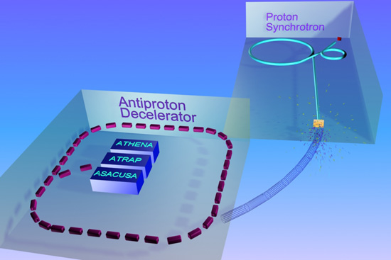
CERN Anti-matter Particle Beam Obliterates Living Cells

For the past three years, scientists at CERN have been experimenting with anti-proton beams and their effect on living cells. They have discovered that anti-proton beams are four times more deadly to living tissue than proton beams. This discovery offers promise of safer and more effective treatments for cancer.

Current particle beam therapy commonly uses protons to destroy tumour cells inside a patient. The ACE experiment directly compared the effectiveness of cell irradiation using protons and antiprotons. To simulate a cross-section of tissue inside a body, tubes were filled with hamster cells suspended in gelatine. Researchers sent a beam of protons or antiprotons with a range of 2 cm depth into one end of the tube, and evaluated the fraction of surviving cells after irradiation along the path of the beam.

The results showed that antiprotons were four times more effective than protons. When comparing a beam of antiprotons with a beam of protons that cause identical damage at the entrance to the target, the experiment found the damage to cells inflicted at the end of the beam path to be four times higher for antiprotons than for protons. Michael Holzscheiter, spokesperson of the ACE experiment, summarises: “To achieve the same level of damage to cells at the target area one needs four times fewer antiprotons than protons. This significantly reduces the damage to the cells along the entrance channel of the beam for antiprotons compared to protons. Due to the antiproton's unsurpassed ability to preserve healthy tissue while causing damage to a specific area, this type of beam could be highly valuable in treating cases of recurring cancer, where this property is vital.”

Antiprotons are antimatter; they have to be produced in small amounts in a laboratory with the help of a particle accelerator. When matter and antimatter particles meet, they annihilate, or destroy each other, transforming their mass into energy. The experiment makes use of this property as the antiproton would annihilate with a part of the nucleus of an atom in a tumour cell. The fragments produced from the energy released by the annihilation would be projected into adjacent tumour cells, which are in turn destroyed.
Source.

Proton beams have been put to good use treating prostate cancer and other cancers. If anti-proton beams are even more effective, with less incidental normal tissue destruction, the new treatment would contribute to both quantity and quality of life.

This is still a case of swatting flies with sledge-hammers, but at least we will be swinging the hammers with a bit more finesse. Newer methods of programming viruses, bacteria, and immune cells to target cancers would seem to be much more potentially accessible methods of cancer treatment for patients all around the world. Even so, it is good to put multi-billion dollar physics facility to the use of saving lives.

Labels:

Bookmark and Share

31 August 2006

Cancer, Hair, Germs, and Steel

First, steel. This story reports on a recently finished bridge that used a new, better form of steel. The copper alloy steel was developed at Northwestern University. The steel has a strength of 70,000 pounds per square inch (psi) compared with 50,000 psi in commonly used structural steel. It is also easy to weld, and tests have shown it has high-impact toughness at low temperatures. In addition, the high copper content gives the alloy much better resistance to atmospheric corrosion than other high-performance steels. Source. I cannot help but wonder why it took 9 years before this better steel was used in large structures when it seems to have so many advantages.

Next, hair. Scientists in the UK have discovered how to command skin cells to become hair follicles. "Which cells are transformed into hair follicles is determined by three proteins that are produced by our genes.

"Our research has identified how one of these proteins working outside of the cell interacts at a molecular level to determine an individual's hair pattern as the embryonic skin spatially organises itself."
Source.
The only treatments likely to earn more money for their developers than the cure for baldness, might be the cures for obesity, a true aphrodisiac, and a true life extension drug.

Third, cancer. Researchers at UCR have added to the knowledge of normal prevention of cancer transformation in cells. Liu published her research findings in a featured article titled Mechanistic insights into maintenance of high p53 acetylation by PTEN, in the Aug. 18 issue of Molecular Cell. Co-authors include UCR colleagues Andrew G. Li, Landon G. Piluso Xin Cai and Gang Wei; with William R. Sellers in the Department of Medical Oncology, Dana Ferber Cancer Institute of Harvard University in Boston.

They found that when a cell’s DNA becomes damaged, PTEN forms a complex with another protein, p300, which in effect, switches on p53, a very important tumor suppressor.

“I would like to continue to expand our understanding of how p53 is activated in conjunction with PTEN and under what circumstances it functions to protect the cell,” Liu said.
Source.

Finally, germs. Bacterial resistance occurs due to routine mutations in microbes, and is a major cause of morbidity and mortality. Scientists at St. Jude Children's Research Hospital have made a finding that may give drug developers a new advantage in the fight against the gram positive pathogens such as strep and staph. Rock's team showed that gram-positive pathogens first use PlsX to synthesize a compound called fatty acyl-phosphate, then use PlsY to transfer the fatty acid to G3P. These steps initiate membrane phospholipid formation required for cell growth.

"Our discovery of PlsX and PlsY not only solved a troublesome mystery," Rock said. "It's also important because identifying the essential components required for disease-causing bacteria to grow and multiply is a key part of developing new strategies for controlling infections."
Source.

Many microbiologists believe that the mutating ability of microbes is so great as to make it unlikely that science will ever develop antibiotics that microbes are unable to resist, eventually. Personally, I believe that evolution only has a limited set of tools for each class of organism. It is up to science to learn what these tools are, then to devise treatments that are not susceptible to those tools. Doing that will require persistence, heightened perception, and invention on the part of scientists.

Update
: For an example of a novel attempt to bypass bacterial resistance, read this newsrelease.

Labels: ,

Bookmark and Share

02 August 2006

Aptamers vs. Antibodies--Early Cancer Detection

Aptamers offer the utility for biotechnological and therapeutic applications as they offer molecular recognition properties that rival that of the commonly used biomolecule, antibodies. In addition to their discriminate recognition, aptamers offer advantages over antibodies as they can be engineered completely in a test tube, are readily produced by chemical synthesis, possess desirable storage properties, and elicit little or no immunogenicity in therapeutic applications. Source.

Aptamers are relatively short molecules (15-50 bases) of RNA, DNA or synthetic analogs that are designed to bind tightly and specifically to target molecules such as proteins. Aptamers can display highly specific binding due to the complex three-dimensional conformations short oligonucleotides can adopt. As these conformations are directly dependent upon primary sequence, synthetic or "genetic evolution" selection techniques can theoretically be used to generate aptamers with almost any specific binding characteristic, especially for any protein.

....Aptamers may someday emerge as an alternative or complement to antibodies as research reagents and diagnostic sensors. It is possible that aptamers may be designed to target proteins for capture and measurement assays, as well as, bind to specific regions of proteins as "agonists" or "antagonists" for functional research. The potential diagnostic use of aptamers for in vivo imaging is also under investigation. However, prior to more widespread use, the technology must be developed to more accurately and efficiently bind specific target proteins and regions.
Source.

Now researchers at the University of Florida have developed aptamers that can diagnose leukemic cells at an early stage, even distinguish between types of cancerous leukocytes:

“Normally, definitive diagnosis of cancer requires a visual examination of the tumor, which is an invasive and time-consuming process,” explained Weihong Tan, a UF professor of chemistry and lead author of the paper. “Most importantly, this process is not suitable for early detection, when the cancer is at its most treatable.”
Clinicians can sometimes use antibodies, proteins that recognize and fight bodily intruders, to identify different types of cancer. That’s the case, for example, with the prostate-specific antigen test for prostate cancer. Antibodies are preferable to diagnosis by appearance because they are consistent and accurate, but they are only available for a selected few cancers, Li said.

Tan, a member of the UF Shands Cancer Center and the UF Genetics Institute, said that scientists know that cancer tissue has a unique molecular fingerprint that can distinguish it from healthy tissue. But attempts to target cells via these fingerprints have largely proved futile because there are few molecular tools to recognize the fingerprints. The UF team sought to create these tools in the form of aptamers, or short strands of chemically synthesized DNA. These aptamers exploit the differences on the surface of cells to discern cancerous ones. Key to the approach is it does not require prior knowledge of cancer indicators, Tan said.

“Using the cell-based aptamer selection strategy, we can generate aptamers which can specifically recognize any kind of cells without prior knowledge of molecular changes associated with the disease,” he said.

In experiments, the researchers showed they could successfully design sets of aptamers that would recognize leukemia cells that had been mixed in with normal bone marrow cells. The aptamers also successfully distinguished leukemia T-cells from lymphoma B-cells. Both results indicate that the aptamer method could be used to identify many different types of cancer, researchers said.

Clinicians using such molecular probes should be able to “find cancer in a much earlier stage when the tumors are much smaller,” enabling doctors to begin treatment earlier, Li said.
Source.

Early diagnosis of cancer cells using aptamers and/or advanced antibodies, can make cancer screening more accurate, and can bring about earlier diagnosis of cancers. Combining earlier diagnosis with safer and more effective non-invasive cancer treatments, will lead to fewer cancer deaths and less traumatic cancer therapies.

Labels: ,

Bookmark and Share

25 July 2006

Bubbles to Kill Cancer ( with a few cruel asides)

University of Michigan Medical School Radiology researchers, are looking into using gas bubbles to embolize small arteries leading to cancerous tumours.

The process of blocking blood flow to a tumor is called embolization, and using gas bubbles is a new technique in embolotherapy. What makes it so promising is that the technique allows doctors to control exactly where the bubbles are formed, so blockage of blood flow to surrounding tissue is minimal, said Joseph Bull, assistant professor of biomedical engineering at U-M.

The research of Bull and collaborator Brian Fowlkes, an associate professor in the Department of Radiology in the U-M Medical School, is currently focused on the fundamental vaporization and transport topics that must first be understood in order to translate this developmental technique to the clinic.

In traditional embolotherapy techniques, the so-called cork that doctors use to block the blood flow—called an emboli—is solid. For instance, it could be a blood clot or a gel of some kind. A major difficulty with these approaches is restricting the emboli to the tumor to minimize destruction of surrounding tissue, without extremely invasive procedures, Bull said. The emboli must be delivered by a catheter placed into the body at the tumor site.

Gas bubbles, on the other hand, allow very precise delivery because their formation can be controlled and directed from the outside, by a focused high intensity ultrasound.

This envisioned technique is actually a two-step process, Bull said. First, a stream of encapsulated superheated perfluorocarbon liquid droplets goes into the body by way of an intravenous injection. The droplets are small enough that they don’t lodge in vessels. Doctors image the droplets with standard ultrasound, and once the droplets reach their destination, scientists hit them with high intensity ultrasound. The ultrasound acts like a pin popping a water balloon. After the shell pops, the perfluorocarbon expands into a gas bubble that is approximately 125 times larger in volume than the droplet
More at the Source. Hat tip Medgadget.

This is a potentially good approach to embolizing arteries. We all know the damage that air emboli do to unwary SCUBA divers who hold their breaths on ascent, or what nitrogen bubbles in the blood do to sufferers of the Bends, when they fail to de-compress adequately. Whether to treat cancer, or to stop aneurysms from bleeding, safe methods of arterial occlusion/embolization have to be developed.

Now for the cruel asides. While looking over the U Mich. news service webpage, I noticed some headlines on the sidebar. The first headline was "Why Money Doesn't Bring Happiness." The second headline read, "Study Reveals a Disproportionately High Number of Minorities and Poor Live near Toxic Waste Facilities".

Now, anyone with an IQ over 90 can read those two headlines and see an inconsistency. So why did the media people at UMich fail to see something that so clearly made them look silly? You can reach your own conclusions.

As for the second headline: " a disproportionately high number of minorities and poor live near toxic waste facilities". Likewise, anyone with an intelligence high enough to get a job begging on the streetcorner can see that using the phrase "minorities and poor" in that sentence is bad technique (although quite PC).

It is obvious to almost everyone able to ambulate to the corner store, that if a minority is wealthy enough, he will choose to live away from a toxic waste facility. Being a minority has nothing to do with it--it is being poor that forces someone to live where he does not want to live, at least in Canada, the US, and the rest of the Anglosphere. So that although money may not bring happiness, it can certainly help you to do away with several obvious causes of unhappiness.

You cannot expect a journalist to understand the concept of epidemiological confounding, or even simple contradiction, but an IQ above 80 and common sense, should lead journalists to think about what they write, far more often than they seem to.

If it was "all about the story", you would think that journalists would try harder to get the story right. But it is really about being a member of the club, and hoping if you say the right things, the club will give you a Pulitzer, or a lucrative job with a high paying publisher. Think about the fact that it is the low achieving students who become journalists (and schoolteachers), and contemplate what that means for the quality of information that filters down to the public and the schools.

Labels:

Bookmark and Share

11 July 2006

Fireflies Help Fight Cancer: Restoring p53 Tumour Suppressor Function

Researchers at the University of Pennsylvania School of Medicine have used the luminescence from fireflies to help screen small molecules that might help restore p53 tumour suppressor gene function to cancer cells that had lost that protection through mutation.

In an attempt to defend the body, a normal p53 protein will bind to DNA during periods of cellular stress or damage. The binding of p53 to DNA initiates downstream reactions that keep the stressed cells from multiplying. Under normal conditions, p53 will activate the p21 gene, causing the cell cycle to freeze, halting cell proliferation; p53 will activate KILLER/DR5, which signals for cell death, or apoptosis. Chemotherapy and radiotherapy set out to deliberately stress tumor cells in hopes of promoting their self-destruction. Unfortunately, mutations to the p53 gene disrupt the intracellular defense system.

"Mutants of p53 that occur in human cancer fail to bind to DNA or to activate target genes, such as p21 and KILLER/DR5," explains El-Deiry, who is also the Co-Program Leader of the Radiation Biology Program at the Abramson Cancer Center at Penn. "Therefore, when cells are stressed or damaged, p53-mutant cells fail to shutdown and continue to divide uncontrollably." The development of a drug screen by El-Deiry's lab allowed the researchers to trace the activity of small molecules in p53-mutant cancer cells.

The small molecule drug screen, developed by El-Deiry's lab, was created by inserting firefly luciferase, a reporter gene capable of emitting light, into human tumor cells carrying the p53 mutation, and observing the subsequent response.

"Just as fireflies emit light that we can see with our eyes, the cancer cells were engineered to emit light if a p53-like response was triggered by any of the small molecules that we examined," explains El-Deiry.

The small molecules screened by El-Deiry's research group were obtained from the Developmental Therapeutics Program at the National Cancer Institute. The molecules represent many classes of compounds and include both natural and man-made chemicals.

"One by one, we introduced the small molecules to the p53 mutant cancer cells, which possessed the luciferase reporter gene and screened for light emissions," describes El-Deiry. The light emissions displayed by the live cell imaging instrumentation revealed which molecules were able to achieve p53 responses in the abnormal cancer cells. Further testing exposed the ability of high doses of several groups of the small molecules to kill human cancer cells in cell culture and in mouse models implanted with human tumors.

"Our work provides a blueprint for how molecularly targeted therapy can be discovered using new optical imaging technology," states El-Deiry. "This is very important going forward in the era of molecular medicine and individualized therapy for cancer patients."
Source.

This is a clever approach to cancer drug discovery. This type of drug, if effective, could become a useful second line defense against cancer, after prevention. As cancer screens become more sensitive--able to pick up malignancies very early--the need for safe broad spectrum anti-tumour drugs such as this type of tumour suppressor gene restorer becomes greater. The key is to find such tumour suppressor gene augmentors/restorers that are both safe and effective. Good luck to the UPenn. lab.

Labels: ,

Bookmark and Share
Older Posts
Al Fin Main Page
Enter your Email


Powered by FeedBlitz
Google
WWW AL FIN

Powered by
Blogger

``