06 December 2012

4 Million Switches to Control 20,000 Genes: An Ongoing Revolution in Biology

We are living on a planet that has been transformed by biology into a birthplace and cradle of proto-intelligent life. The biological transformation of our world is still at a very early stage. The true wonders of our ongoing biological revolution have barely been hinted at.
When the ENCODE Project announced that so-called "junk DNA" actually contains millions of gene control switches to control roughly 20,000 genes, the educated public was suddenly made aware of something that working biologists have known for decades: We humans are not in Kansas anymore, Toto.
The human genome is packed with at least 4 million gene switches that reside in bits of DNA that once were dismissed as junk but that turn out to play critical roles in controlling how cells, organs, and other tissues behave. The discovery, considered a major medical and scientific breakthrough, has enormous implications for human health because many complex diseases appear to be caused by tiny changes in hundreds of gene switches.

The findings are the fruit of an immense federal project, involving 440 scientists from 32 global labs. As they delved into the junk — parts of the DNA that are not actual genes containing instructions for proteins — they discovered it is not junk at all. At least 80 percent of it is active and needed. _BG

Protein Transcription Factors: One of Many Factors in Gene Switching

Just when the educated public thought it was beginning to understand how cells work, they are told that the mechanisms of life are orders of magnitude more complex than they previously believed.

The secret to complex life is not just the mechanisms of DNA transcription to RNA, and RNA tranlation to proteins. Complex life is an astounding swirl of circular logic and control circuits of cell signaling. Some genes are constantly being switched on and off, while other genes are silenced permanently or over long periods of time.

But we are discovering ways to alter the natural order of cell signaling and gene switching -- and that ability to change the natural scheme of things amounts to a building revolution in our biological world.

Here is a quick example of a discovery in cell switching which may lead to the ability to quickly repair damage to heart muscle from hear attacks:
MicroRNAs are short segments of RNA whose purpose is to cause genes to switch on and off. To find out which ones are responsible for causing heart cells to divide, the team studied 875 of them taken from a human heart and implanted into rodent muscle. In so doing they found 204 of them that reactivated cell proliferation and 40 and that did so strongly. They then chose the two strongest and injected them into the hearts of live mice that had been caused to suffer damage to their hearts, using a harmless virus as a carrier.

After two weeks, the mice that had been injected with the MicroRNAs showed less damage than prior to the treatment, indicating regeneration had occurred. After two months, the damaged tissue area had been reduced by half. The team also noted that contraction strength improved as did other heart functions that were measured.

The research team concludes by suggesting that their method of using MicroRNAs to induce regeneration of damaged heart tissue might be used someday soon to treat heart attack victims... _MXP
Abstract of study in Nature

Heart disease is the primary cause of death in most developed countries. The ability to rapidly heal heart muscle damage after heart attacks would likely prolong the productive lives of hundreds of thousands of people in the developed world every year.

Cell switching effects of micro RNA and Transcription Factor networks (PDF)

When we consider a world where humans have achieved the mastery of cell signaling and gene switching, we are not necessarily looking at a world of immortal, universally brilliant, and physically powerful humans. We should look at these things in relative terms, rather than in absolutes. Compared to monkeys, humans are longer-lived and quite capable in a broader range of activities and environments.

Likewise, compared to modern humans, those future people who have achieved mastery over biology will live longer lives, and possess a significantly broader range of aptitudes and capabilities over a greater number of environments.

Biology has its shortcomings, of course. We are likely to discover ways of bypassing and substituting for, much of the evolved complexity of biological cells, organs, and organisms for the sake of improved reliability.

But that will have to be done in a carefully considered and cautious manner. We have to be sure that we do not sacrifice too much resiliency for the sake of reliability within a narrow niche of functioning. No one wants to be a Dodo bird.

Labels: , , ,

Bookmark and Share

12 January 2010

Test Bed for Radical Evolution of Cells

Harvard researchers have devised a novel cell culture platform, which sets the stage for a program of radical designed cellular evolution. The test bed (of nails) provides a surface for cells to grow, but also provide a ready means of inserting a wide array of molecular, genetic, or biological (viruses) elements into the growing cellular machinery. Scientists can then observe the effects of various inserted elements upon the growth and behaviour of the cells.
Author Hongkun Park, a professor of chemistry and physics at Harvard University, says that, in theory, "you can put more or less any molecule in more or less any kind of cell." If the method proves effective, it could greatly speed the ability to manipulate cells in a variety of applications, including stem-cell reprogramming and drug screening.

Park's lab recently discovered that cells can be grown on beds of vertical silicon nanowires without apparent damage to the cells. The cells sink into the nanowires and within an hour are impaled by the tiny spikes. Even resting on this bed of needles, cells continue to grow and divide normally. This setup makes it possible to directly interface with the cell's interior through the nanowires. "Since we now have direct physical access, we can deliver molecules into cells without the restrictions of other techniques that are available," Park says. He adds that while his lab has found that many different types of cells seem to accommodate the tiny wires without negative effects, further studies will be needed to examine whether any important cell behaviors are affected.

To use the nanowires to deliver molecules, Park's team first treated them with a chemical that would allow molecules to bind relatively weakly to the surface of the nanowires, then coated the wires with a molecule or combination of molecules of interest. When cells are impaled on the nanowires, the molecules are released into the cells' interior. The chemical treatment of the wires could potentially be manipulated to control the binding and release of molecules--releasing them more slowly, for instance--and the wires can be constructed at different lengths to reach different parts of the cell. To demonstrate the method's flexibility, the team used the approach to deliver chemicals, small RNA molecules, DNA, and proteins into a range of cell types.

The beds of nanowires can be arranged on microarrays suitable for rapid experiments and imaging cells under a microscope. These microarrays can be "printed" with different patterns or combinations of molecules, making it possible to test many different molecules at once on an array of cells. The authors believe it could be possible to screen 20,000 different proteins or other chemicals on cells within a single microscopic slide.

...Thorsten Schlaeger, a stem-cell researcher at Children's Hospital Boston, is investigating the potential of the approach for reprogramming stem cells. His lab is interested in turning embryonic and induced pluripotent stem cells into blood stem cells like those found in the bone marrow. Currently, this task requires infecting cells with a virus to introduce new genes into their DNA, and, Schlaeger says, "there's no good alternative right now." Schlaeger's team is looking for better ways to manipulate cells, as well as ways to screen stem cells for factors that can transform them from one cell type to another. "It's hard to say what will be possible because it's new, but it's intriguing," he says._TechnologyReview

Early applications of the technology will include micro-arrays for high-throughput drug testing, and other rapid screening applications. But the technology virtually screams "rapid evolution"!

Imagine growing complex neural networks onto such test beds -- which are equipped to inject and extract both a wide range of molecular materials, but also various types of electromagnetic stimuli. The idea would be to grow entire cortical columns on a 3 dimensional implementation, then to connect the cultured cortical columns together one at a time -- all the while closely monitoring every aspect of the chemical, genetic, and electrophysiological functioning of the complex networks.

That is just the beginning. And what a simple idea.

Labels: ,

Bookmark and Share

14 December 2009

Life: Isn't That Like Magic?

To human minds, the molecular mechanisms of life are very much like magic.  Things happen so quickly on the molecular level -- and so far out of human sight -- that by the time we understand what is happening we are often thrown completely off our stride.

We know that an animal's genome changes over time, but we have little idea how animals change and evolve.  Indiana University and University of New Hampshire biologists have made some important discoveries regarding evolution driven by intron formation inside genetic sequences.   Introns are non-coding DNA sequences "inside" genes, that make up much of DNA in cells.   As different introns are inserted into genes of a species, members of the species may begin behaving differently -- genetically speaking.  This discovery could prove extremely important.

Speaking of introns, the cell needs to "edit out" all the mRNA that does not code for a gene's protein, in order to translate only the gene's "message" into protein (not the intron's).   Tel Aviv University researchers have made some pivotal discoveries in how RNA is edited, based upon the way it is transcribed from the DNA.  Their work may have important implications for cancer research as well as basic research in gene expression.

Johns Hopkins University scientists have learned more about particular epigenetic processes that seem to play a special part in an organism's ability to adapt to its environment, in evolution.   The research looks at patterms of methylation of genes, which can have the effect of "randomizing" the organism's response to the environment for different members of the same species -- for example, modifying size, shape, strength, skin tone, disease resistance, etc.   Another way of modifying (in fact, randomizing) gene expression -- methylation, epigenetics.

Korean researchers have learned more about a "growth regulationg" micro-RNA in fruit flies, miR-8, which plays a significant role in determining the animal's size.  Humans have a similar micro-RNA referred to as miR-200, which seems to affect a person's size and weight via affects on insulin.   Yet another gene expression modifier -- micro RNA.

Baylor College of Medicine researchers are looking into "master gene" Math1 , which seems to help coordinate the different nerve centers for hearing, balance, proprioception (position and orientation of body parts), and interoception (the detection of internal body states such as a full bladder).  It has a lot to do with why you can get up in the middle of the night -- half asleep -- and navigate to the bathroom and back, without causing injury to yourself and damage to your household.   Can you believe it?  A gene that controls and coordinates conscious / unconscious behaviour?

McGill University scientists are looking at a protein that influences DNA shape  and gene expression -- helicase protein translation initiator DHX29.  This protein is important in regulating protein synthesis, and cell proliferation.  It is associated with cancer cell growth -- the less DHX29, the less cancer cell growth.  Yet another form of gene expression:  helicase protein translation initiators.

UCSD researchers in La Jolla are studying how atypical anti-psychotic drugs such as olanzepine and clozapine are able to improve a schizophrenic's cognitive function enough to sometimes go back to work and be productive.  Using ingenious bio-sensor "sniffers" they call CNiFERs, they were able to determine that atypical anti-psychotics have a strong blocking effect on the M1 (muscarinic 1) acetylcholine receptor in rats.  They hope to modify their CNiFERs to "spy on" other receptors in pursuing this important research in cell signaling.

You may begin to understand that there is no separation between the molecular / genetic level and the organismic / behavioural level of animals.  That is why it is so important for us to understand the molecular nature of the animal -- because it underlies everything else.

Labels: , ,

Bookmark and Share

05 May 2009

Pin-Point Precision: Nano-Cell Biology

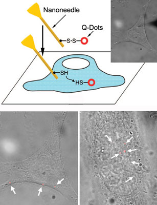
These new gold-plated boron nitride nanotube - nanoneedles from the University of Illinois are a good example of technology convergence. The ability to discover the intimate workings between cell and molecular biology have never been as strong, thanks to the use of these special nano-biotechnological devices. Here is how it works:
To create a nanoneedle, the researchers begin with a rigid but resilient boron-nitride nanotube. The nanotube is then attached to one end of a glass pipette for easy handling, and coated with a thin layer of gold. Molecular cargo is then attached to the gold surface via “linker” molecules. When placed in a cell’s cytoplasm or nucleus, the bonds with the linker molecules break, freeing the cargo.
With a diameter of approximately 50 nanometers, the nanoneedle introduces minimal intrusiveness in penetrating cell membranes and accessing the interiors of live cells.

The delivery process can be precisely controlled, monitored and recorded – goals that have not been achieved in prior studies. “The nanoneedle provides a mechanism by which we can quantitatively examine biological processes occurring within a cell’s nucleus or cytoplasm,” said Yang Xiang, a professor of molecular and integrative physiology and a co-author of the paper. “By studying how individual proteins and molecules of DNA or RNA mobilize, we can better understand how the system functions as a whole.” _Nanowerk
Extremely cool tool. The possibilities are beyond current comprehension. These techniques will no doubt be used in induced stem cell studies, and in all kinds of differentiation - dedifferentiation studies. And that is only the beginning.

The dream is consilience -- the unified study of knowledge in real time. This type of tool allows for one small part of the dream.

Labels: ,

Bookmark and Share

02 December 2007

Probing the Cell With Many Fingers

Eric Drexler talks about nanobots that roam the human body, looking for problems in cells--then jumping into the cell to repair any damage or dysfunction. Developing these "cell repair machines" will take some time. In the meantime, we need to learn different ways to probe the inner and outer workings of cells.

For example, we may want to develop a tiny nano-voltmeter to monitor the internal electric field of cells:
"The basic idea behind this field of research is to follow cellular processes---both normal and abnormal---by monitoring physical properties inside the cell. There's a long history of research on the chemistry happening inside the cell, but now we're getting interested in measuring the physical properties, because physical and chemical processes are related," said Kopelman, who is the Richard Smalley Distinguished University Professor of Chemistry, Physics and Applied Physics.

With a diameter of about 30 nanometers, the spherical device is 1,000-fold smaller than existing voltmeters, Kopelman said. It is a photonic instrument, meaning that it uses light to do its work, rather than the electrons that electronic devices employ.

Kopelman's former postdoctoral fellow Katherine Tyner, now at the U.S. Food and Drug Administration, used the nano-voltmeter to measure electric fields deep inside a cell---a feat that until now was impossible. Scientists have measured electric fields in the membranes that surround cells, but not in the interior, Kopelman said.

With the new approach, the researchers don't simply insert a single voltmeter; they're able to deploy thousands of voltmeters at once, spread throughout the cell. Each unit is a single nano-particle that contains voltage-sensitive dyes. When stimulated with blue light, the dyes emit red and green light, and the ratio of red to green corresponds to the strength of the electric field in the area of interest.
Eurekalert

Clever, no? Or perhaps researchers could use X-rays to take snapshots of the cell in action:
A research team led by Jue Chen, an associate professor of biological sciences, obtained a snapshot of the tiny protein gate complex that opens and closes pathways through the protective cellular membrane. The gates, operated by small protein machines that push them open and closed, bring nutrients into the cell and flush out waste.

The Purdue-led team was the first to achieve an image of the middle step of the process, capturing the molecular interactions as material passes through the membrane.

...The research team used X-ray crystallography to obtain a picture of a special protein, called an ABC transporter protein, as it moved material through the cellular membrane. The work was published in last week's issue of Nature.
Purdue

But there are many more approaches (many fingers) used to probe cells:

Etc.

Labels: , ,

Bookmark and Share

04 October 2007

Portable Cell Cultures to Portable Embryo Incubator to Portable Artificial Womb

A recent story about the grandmother in Brasil who gave birth to her own twin grandchildren suggests that there would be a strong demand for an artificial womb, should such a thing ever be perfected. This grandmother was apparently happy to perform this vital service for her daughter. After all, she was able to walk away from the primary responsibilities of childraising when she left the hospital. But how would the dynamics of the same situation have changed in the context of safe and efficacious artificial wombs?

You may remember the microfluidic chip developed by Teruo Fujii of the University of Tokyo, designed to nourish an embryo in its early stages of development before final transplantation into a human womb. And the recent microfluidic cell culture incubator developed at Johns Hopkins is an impressive development along the same lines.
In a recent edition of the journal IEEE Transactions on Biomedical Circuits and Systems, the Johns Hopkins researchers reported that they had successfully used the micro-incubator to culture baby hamster kidney cells over a three-day period. They said their system represents a significant advance over traditional incubation equipment that has been used in biology labs for the past 100 years.

...In contrast, the thumb-size system developed by the Johns Hopkins engineers is self-contained and requires no external heating source. A drop of liquid containing living cells is injected into a port and flows through one of the microfluidic channels. A nutrient solution — the cells’ food – is also added in this manner.

The cells gravitate toward and stick to the surface of the microchip. The chip contains a simple heating unit – a miniature version of the type found in a common toaster – and is equipped with a sensor that continually checks to make sure the proper temperature is maintained. For human cells, this is usually 37 degrees Celsius or 98.6 degrees Fahrenheit. The chip is connected to a computer that controls the sensing and heating process. The prototype is connected to a computer via a hard wire, but the inventors say a wireless version would be the next step.

A gas-permeable membrane on the incubator allows the microsystem to exchange carbon dioxide and oxygen but keeps out bacteria that could contaminate the cell culture. If a cell colony grows too large, an enzyme can be injected into one of the microfluidic ports to detach and flush away surplus cells without destroying the primary cell culture.
Source

Quite clever indeed. From such microfluidic culture devices, it is not such a stretch to imagine a staged device with graduated chambers (or a single expandable chamber) that accepts an IVF embryo at one end, and nine months later delivers a fully developed neonate out the other.

Bioethicists will no doubt agonize over the concept from now until the next millenium, but the fact is there is a demand for such devices. Where there is a demand, human ingenuity will usually created a supply.

A previous Al Fin article discussed two of the most famous would-be developers of artificial wombs, scientists Kuwabara in Tokyo, and Liu at the Center for Reproductive Medicine and Infertility. Other researchers are working in the background, developing the necessary techniques, devices, and software that would allow a single cell to develop into a fully developed neonate in vitro.

One step at a time, largely unnoticed by the public. That is how most science advances.

Labels: ,

Bookmark and Share

31 August 2007

Building a Brain--One Neuron at a Time

Neuroscientists do not really understand individual neurons very well. They are only now just learning how to culture and study neurons at low density, without contaminating them with serum or blood plasma.
First, the researchers scaled down the size of the fluid-filled chambers used to hold the cells. Chemistry graduate student Matthew Stewart made the small chambers out of a molded gel of polydimethylsiloxane (PDMS). The reduced chamber size also reduced – by several orders of magnitude – the amount of fluid around the cells, said Biotechnology Center director Jonathan Sweedler, an author on the study. This “miniaturization of experimental architectures” will make it easier to identify and measure the substances released by the cells, because these “releasates” are less dilute.

...Second, the researchers increased the purity of the material used to form the chambers. Cell and developmental biology graduate student Larry Millet exposed the PDMS to a series of chemical baths to extract impurities that were killing the cells.

Millet also developed a method for gradually perfusing the neurons with serum-free media, a technique that resupplies depleted nutrients and removes cellular waste products. The perfusion technique also allows the researchers to collect and analyze other cellular secretions – a key to identifying the biochemical contributions of individual cells.


... This combination of techniques enabled the research team to grow postnatal primary hippocampal neurons from rats for up to 11 days at extremely low densities. Prior to this work, cultured neurons in closed-channel devices made of untreated, native PDMS remained viable for two days at best.

The cultured neurons also developed more axons and dendrites, the neural tendrils that communicate with other cells, than those grown at low densities with conventional techniques, Gillette said.

The technique is described this month in the journal of the Royal Society of Chemistry – Lab on a Chip.
Source

Neural reductionism at its best, eh? By culturing individual neurons and small groups of neurons, and learning how to supply the needed growth factors and nutrients without potentially contaminating plasmas and serums, neuroscientists can begin to understand these cells at a very basic level. Then they can learn to combine them, along with their support (glial) cells. Eventually they can add capillaries, lymph vessels, etc. and move up from there. By understanding each level of complexity as they develop, neuroscientists can better envision the combinatorial possibilities.

One of the more intriguing uses to which these micro-cultures can be put, is brain-machine interfaces. By learning to develop neurons and small groups of neurone (micro-nets) away from normal biological substrate (brains, blood, and tissue fluid), it is only a short step to micro-channel support chips that can function as interfaces to machines. Neurophilosopher discusses a related development.

This is an important development. Together with improved techniques for manipulating neuronal stem cells and progenitors, this development points directly to better "wet" neural net models of brain, and better nerve-machine interfaces.

Labels: ,

Bookmark and Share

30 June 2007

Bacterial Genome Transplanted, Next Step--Synthetic Organism?

After replacing DNA in mycoplasma capricolum organism with the chromosome from mycoplasma mycoides, scientists at the Ventner Institute are planning work on a completely synthetic organism.
Scientists at the J. Craig Venter Institute (JCVI), a genomics research facility, transplanted a bacterial chromosome from one type of bacteria into another, and have completely replaced an entire bacterial genome and its expression. The work of Carole Lartigue, Ph.D. and colleagues was published in the latest issue of Science:

The JCVI team devised several key steps to enable the genome transplantation. First, an antibiotic selectable marker gene was added to the M. mycoides LC chromosome to allow for selection of living cells containing the transplanted chromosome. Then the team purified the DNA or chromosome from M. mycoides LC so that it was free from proteins (called naked DNA). This M. mycoides LC chromosome was then transplanted into the M. capricolum cells. After several rounds of cell division, the recipient M. capricolum chromosome disappeared having been replaced by the donor M. mycoides LC chromosome, and the M. capricolum cells took on all the phenotypic characteristics of M. mycoides LC cells.

As a test of the success of the genome transplantation, the team used two methods -- 2D gel electrophoresis and protein sequencing, to prove that all the expressed proteins were now the ones coded for by the M. mycoides LC chromosome. Two sets of antibodies that bound specifically to cell surface proteins from each cell were reacted with transplant cells, to demonstrate that the membrane proteins switch to those dictated by the transplanted chromosome not the recipient cell chromosome. The new, transformed organisms show up as bright blue colonies in images of blots probed with M. mycoides LC specific antibody.

The group chose to work with these species of mycoplasmas for several reasons -- the small genomes of these organisms which make them easier to work with, their lack of cell walls, and the team's experience and expertise with mycoplasmas. The mycoplasmas used in the transplantation experiment are also relatively fast growing, allowing the team to ascertain success of the transplantation sooner than with other species of mycoplasmas.

According to Dr. Lartigue, "While we are excited by the results of our research, we are continuing to perfect and refine our techniques and methods as we move to the next phases and prepare to develop a fully synthetic chromosome."
Source

Synthetic biology is one of many approaches to studying the mechanisms of life. Craig Ventner says that he will create an organism that will solve the energy crisis. Perhaps he will. As long as western civilisation survives the onslaughts of anti-enlightenment thinking, I suspect that organisms that can produce unlimited energy will be the least of achievements from synthetic biology, nano-biology, biologic computing etc.

Because western educational systems do not teach students to use their broad intellectual capacities, most humans--even in the developed world--do not have a clue about the multiple revolutions in scientific discovery that are teetering on the very brink of the activation energy hump. Some students of the singularity believe that the true revolution will require the creation of a friendly superhuman machine intelligence.

Personally, I believe that machine augmentation of human intelligence will be enough--once humans learn to use the intellects they possess. But since the educational establishments are incapable of helping humans learn about their intrinsic capacity, there may be some delay.

Labels: , , , ,

Bookmark and Share

22 June 2007

DNA "Computer" Works Inside Living Cells, Suggests Possibilities

Although DNA computers have been made that can play simple games like tic-tac-toe, programming DNA computers to work inside living cells is much more interesting.
The goal is to inject human cells with DNA that can determine whether a cell is cancerous or otherwise diseased, based solely on the mix of molecules inside the cell. Sensing disease, the DNA might trigger a pinpoint dose of treatment in response. That technology, however, is a long way off. For now, researchers are testing different ways of turning DNA into versatile computers that can detect certain combinations of molecules and respond by producing other molecules.

...RNAi is something that cells do naturally. Cells produce what are known as short interfering RNA (siRNA) molecules, which recognize corresponding DNA sequences in genes and cause them to shut down.

Benenson and colleagues engineered a target gene to be sensitive to several different siRNAs of their own design. In the simplest case, they introduced a single siRNA molecule to switch off a target gene that encoded a fluorescent protein. In more complex cases, a pair of siRNAs or either of two siRNAs switched off another target gene, which in turn switched off a gene for a fluorescent protein. To make sure the system worked as intended, the researchers based their siRNAs on those of other species, they report in a paper published online today by Nature Biotechnology.

In principle, the RNAi technique can reach great heights of complexity, Benenson says, by making genes sensitive to more and more siRNAs in various combinations. "The scalability is very important, because eventually you want to make complex decisions," he says.

He says the next step is figuring out how to make the molecules inside a cell—such as those that are overproduced in cancer—trigger the production of siRNAs.
Source

This is a very simple approach to DNA "computing", but for all its conceptual simplicity it suggests possibilities that are much more complex. It is best to go very slowly and carefully. The type of control of gene expression hinted at here is not only promising as a cure for cancers, it is threatening.

This type of research appears ideal for a synthetic biological organism. At this time synthetic biologists are attempting to design the simplest possible cells, from other species, by including the smallest possible gene set for viability. Presumably, as the synth biologists master the simpler life forms, they will attempt to create more complex organisms.

Synthetic organisms could potentially become ideal biological models for studying various human diseases. Eventually, synthetic organisms and biological systems could replace most animal models--eliminating much of the need for animal research and testing.

This is yet another research field that bears close watching.

Labels: , ,

Bookmark and Share

04 June 2007

Artificial Cells, Synthetic Biology, Revolutionary Life Forms

Living cells are able to thrive miles below the earth and the sea, and can survive transit through the vacuum of space. While we are waiting for nanotechnology to come of age, some revolutionary biologists are learning to design novel life forms almost from scratch--life designed for specific human purposes.
The people who are defying Nature's monopoly on creation are a loose collection of engineers, computer scientists, physicists and chemists who look at life quite differently than traditional biologists do. Harvard professor George Church wants "to do for biology what Intel does for electronics"—namely, making biological parts that can be assembled into organisms, which in turn can perform any imaginable biological activity. Jay Keasling at UC Berkeley received $42 million from Bill Gates to create living microfactories that manufacture a powerful antimalaria agent. And then there's Craig Venter, the legendary biotech entrepreneur who made his name by decoding the human genome for a tenth of the predicted cost and in a tenth of the predicted time. Venter has put tens of millions of dollars of his own money into Synthetic Genomics, a start-up, to make artificial organisms that convert sunlight into biofuel, with minimal environmental impact and zero net release of greenhouse gases. These organisms, he says, will "replace the petrochemical industry, most food, clean energy and bioremediation."
Source

Many of the things that Drexler, Merkel, and Freitas expect out of future nanotechnology can be accomplished in the nearer term by specially designed life-forms. Biology has an evolutionary head start of billions of years over nanotech. While Crick and Watson made their earthshaking discoveries in the mid-20th century, equivalent foundational discoveries for workable molecular assembly nanotech have yet to be made.

It is certainly true that a mastery of biology possessed by malevolent individuals could endanger all life on earth, it is more likely that such a mastery would be used to move industrial and chemical processes to cleaner and more sustainable forms.

Labels: , ,

Bookmark and Share
Older Posts
Al Fin Main Page
Enter your Email


Powered by FeedBlitz
Google
WWW AL FIN

Powered by
Blogger

``