02 November 2012

Mind the Man Behind the Curtain


To most people, their awareness of the world seems integrated and generally effortless, automatic. But on deeper levels, billions of syncopated actions are taking places. On yet deeper levels . . . well, we won't go that deep today.
...we have evolved asymmetrical brains, the right and the left hemispheres, separated (as well as connected) by the corpus callosum, such that these two parts of our brains experience the world in very different yet equally necessary ways. _Philosophy and the 2-Sided Brain

Typical Specialisation of Cerebral Hemispheres

The two hemispheres of the cerebral cortex tend to specialise and divide different functions and specialties between them. The ability to use and understand language is usually the province of the left cerebral hemisphere. This means, for example, that if language information is presented to the right hemisphere, that information must be sent across to the left side for interpretation and analysis.

Advanced brain imaging tools combined with state of the art mathematical / computational analysis tools, have allowed researchers to better understand how information is shared between the two sides of the brain.
In the new study, researchers used magnetoencephalography, or MEG, a non-invasive tool that measures the continuous brain activity of nine healthy young adults. The participants were briefly flashed a stimulus that was either a real word or a "word-like" non-word. The stimulus was randomly shown either to the left or right brain, using a split-visual field experimental technique. MEG allows researchers to peer into brain activity at the rate of 512 images per second, while fMRI only allows for images to be recorded at the rate of one image every two seconds.

The researchers processed this temporal data using novel network analysis techniques drawn from the mathematical and physical sciences that enable them to examine the dynamic nature of brain communication. This novel network analysis allows the data to be presented in movie form, rather than as snapshots. The researchers found that the strength of connections between the left and right hemispheres depended on whether the stimulus was shown to the left or right brain hemisphere.

"A larger number of statistically significant connections were present at several time points after the stimulus when the word or pronounceable non-word was shown to the right brain and needed to be transferred to the language-specialized left brain," said Danielle S. Bassett, UC Santa Barbara Sage Junior Research Fellow in the Department of Physics and the Department of Psychological and Brain Sciences.

"The results further suggest that brain function measured with MEG can be broken into functional communities that rearrange their organization over time –– based on whether the left or right brain saw the stimulus, and thus whether inter-hemispheric information transfer must take place," said Bassett._ME
Research paper abstract from PNAS

Abstract of earlier research

Description of brain imaging tools used in this type of research

The man behind the curtain is your brain, and how it works. You may not care about how your brain works, but a lot of other people do. They want to know what makes you tick, even if you could not care less.

Because too often, dancing to another person's tune is what happens to you while you are making other plans.

Labels:

Bookmark and Share

14 September 2012

We Are All Cyborgs Now: Brain Implants that Think

A collaborative team of US scientists have developed a neural prosthesis which seems to exhibit the ability to restore lost cognitive functions -- and even to improve the function above the normal ability.

The study was performed in monkeys, but the findings will almost certainly apply to human brains, with the proper safeguards and modifications.

More from Technology Review:
The researchers used an array of electrodes to record the electrical activity of neurons in the prefrontal cortex of monkeys while they performed a memory task. The prefrontal cortex is involved in decision making and directs many types of cognitive responses associated with memory or other types of information processing.

The five monkeys in the study were trained to play a matching game in which they were shown an image on a screen and then had to use hand movements to steer a cursor to that same image out of two to seven others that they were shown anywhere from one to 90 seconds later.

...From their recordings in the prefrontal cortex, the research team extrapolated a mathematical model of the electrical activity of neurons involved in the movement decision. The study authors had previously shown that this kind of mathematical model, called MIMO—short for multi-input/multi-output—could interpret and replace memories in rats with the neural implant (see "A First Step Toward a Prosthesis for Memory").

In the new study, the model took multiple signals produced by the brain layer that integrates sensory information related to the task. It then extracted the relevant information to choose a particular movement. The implant can stimulate neurons in order to influence the decision to move the hand to select the correct image.

To test the implant's ability to improve or recover the decision process, the researchers gave the monkeys cocaine intravenously, since cocaine disrupts decision making. Without the activity of the implant, cocaine-affected monkeys frequently could not choose the correct image. But with the device, their decision making was on par, if not slightly better, than normal, even under the influence of cocaine. _TechnologyReview

This research represents the early stages of a neuroscientific campaign to gain the ability to replace damaged brain tissue after trauma, stroke, tumours, and abscesses -- as well as the ability to compensate for non-functioning and atrophied tissue from degenerative diseases such as Alzheimer's and Parkinson's.

It will be decades more before such prostheses are able to restore brain damaged persons to normal function -- or to convert normal brains into super-brains. But even marginal and incremental improvement can make a huge difference in the lives of those with brain impairment -- and in the lives of their families and caretakers.

Study Abstract

Labels: , ,

Bookmark and Share

05 September 2012

Posteromedial Cortex: Pathway to the Self?


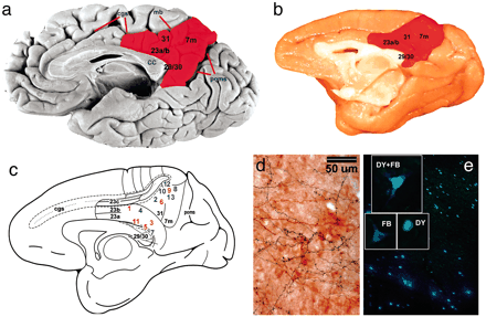
One of the interesting aspects of human consciousness is the sense of the "autobiographical self," which provides us with a sense of personal identity and continuity. Recent research at Stanford U. School of Medicine adds new pieces to the human consciousness puzzle, which cognitive scientists are attempting to assemble. They discovered that the posteromedial cortex was extremely active when recalling events in the person's life, but that this activity was shut down immediately when the patient was asked to do a simple arithmetic problem.

The posteromedial cortex is tucked so deeply into the brain that it is often difficult to examine its ongoing function, using traditional imaging tools such s fMRI. In the Stanford study, researchers took advantage of a previously scheduled neurosurgical procedure being done in a group of epileptic patients, using intracranial electrode monitoring of the PMC.

In a study published online Sept. 3 in Proceedings of the National Academy of Sciences, Parvizi and his Stanford colleagues found a way to directly and sensitively record the output from this ordinarily anatomically inaccessible site in human subjects. By doing so, the researchers learned that particular clusters of nerve cells in the PMC that are most active when you are recalling details of your own past are strongly suppressed when you are performing mathematical calculations.

...The researchers took advantage of a procedure performed on patients who were being evaluated for brain surgery at the Stanford Epilepsy Monitoring Unit, associated with Stanford University Medical Center. These patients were unresponsive to drug therapy and, as a result, suffered continuing seizures. The procedure involves temporarily removing small sections of a patient’s skull, placing a thin plastic film containing electrodes onto the surface of the brain near the suspected point of origin of that patient’s seizure (the location is unique to each patient), and then monitoring electrical activity in that region for five to seven days — all of it spent in a hospital bed. Once the epilepsy team identifies the point of origin of any seizures that occurred during that time, surgeons can precisely excise a small piece of tissue at that position, effectively breaking the vicious cycle of brain-wave amplification that is a seizure.

...The experimenters found eight patients whose seizures were believed to be originating somewhere near the brain’s midline and who, therefore, had had electrode packets placed in the crevasse dividing the hemispheres. (The brain’s two hemispheres are spaced far enough apart to slip an electrode packet between them without incurring damage.)

...Significant portions of the PMC that were “tapped” by electrodes became activated during self-episodic memory processing, confirming the PMC’s strong role in recall of one’s past experiences. Interestingly, true/false statements involving less specifically narrative recall — such as, “I eat a lot of fruit” — induced relatively little activity. “Self-judgment” statements — such as, “I am attractive” — elicited none at all. Moreover, whether a volunteer judged a statement to be true or false made no difference with respect to the intensity, location or duration of electrical activity in activated PMC circuits.

This suggests, both Parvizi and Foster said, that the PMC is not the brain’s “center of self-consciousness” as some have proposed, but is more specifically engaged in constructing autobiographical narrative scenes, as occurs in recall or imagination.

Foster, Dastjerdi and Parvizi also found that the PMC circuitry activated by a recall task took close to a half-second to fire up, ruling out the possibility that this circuitry’s true role was in reading or making sense of the sentence on the screen. (These two activities are typically completed within the first one-fifth of a second or so.) Once activated, these circuits remained active for a full second.

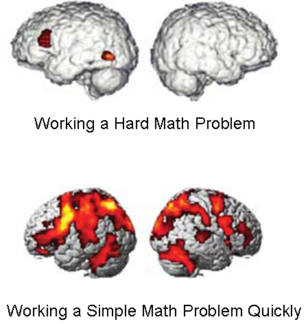
Yet all the electrodes that lit up during the self-episodic condition were conspicuously deactivated during arithmetic calculation. In fact, the circuits being monitored by these electrodes were not merely passively silent, but actively suppressed, said Parvizi. “The more a circuit is activated during autobiographical recall, the more it is suppressed during math. It’s essentially impossible to do both at once.” _Parvizi _via SD
As the researchers suggest, the PMC is not likely to be "the centre of self-consciousness." Rather, the PMC is one part of the human brain's mechanism of self-consciousness -- and a crucial part at that.

It is usually a mistake to claim that a specific part of the brain is "the centre" for a specific cognitive or behavioural function. Rather, different brain modules are integrally tied to various behavioural and cognitive functions, and work in concert to perform cognitive and behavioural functions and actions.

PNAS Neural Connections of PMC in Macaque

PLoS One: Functional Connections of the Human PMC

More: CUNY researchers have discovered that androgen receptors in the visual cortex appear to affect how males see the world through their eyes, as opposed to the way that females see the world. Since androgen receptors are scattered throughout the human cerebral cortex, it is likely that male brains not only see the world differently (statistically), but also perceive the world differently in all of their senses, and feel driven to react to what they perceive in a different manner (statistically) than do females.

Labels: , , ,

Bookmark and Share

10 August 2012

Steps to Better Human Brains

The main obstacle to a more abundant human future is the relatively poor quality of the average human brain. If only we could grow better human brains, and somehow make existing brains work better. Scripps Institute researchers may have discovered one piece of the puzzle, in the longer quest to the development of better human brains.
In mammals, the cortex is made up of six distinct anatomic layers holding different types of excitatory neurons. They are not the uniform layers of a cake, but rather, they are more like the layers wrapped around an onion. The smaller lower layers, on the inside, host neurons that connect to the brain stem and spinal cord to help regulate essential functions such as breathing and movement. The larger upper layers, closer to the outer surface of the brain, contain neurons that integrate information coming in from the senses and connect across the two halves of the brain.

The upper layers are a "relatively young invention," evolutionarily speaking, having been greatly expanded during primate evolution, said Mueller. They give humans in particular the unique abilities to think abstractly, plan for the future and problem-solve.

Previously, it was thought that all cortical neurons -- those making up both the lower and upper layers -- came from the same type of stem cell, called a radial glial cell, or RGC. A neuron's fate was thought to be determined by the timing of its birth date. The Scripps Research team, however, showed that there is a distinct stem cell progenitor that gives rise to upper layer neurons, regardless of birth date or place.

...Published in the August 10, 2012 issue of the journal Science, the new research reveals how neurons in the uppermost layers of the cerebral cortex form during embryonic brain development. _SD

They are saying that they think they have discovered a type of stem cell which gives birth to upper layer neurons, which seem to constitute one important difference between higher mammalian brains in primates, and lower mammalian brains, in neuroarchitecture.

More intriguing, they think the stem cell will migrate into the upper cortical layers regardless of when it is introduced. Would you like more upper layer neurons?
Cerebral Cortical Layering

Another fascinating bit of brain research comes from MIT, recently published in Nature:
There are hundreds of different types of neuron in the brain; most are excitatory, while a smaller fraction are inhibitory. All sensory processing and cognitive function arises from the delicate balance between these two influences. Imbalances in excitation and inhibition have been associated with schizophrenia and autism.

"There is growing evidence that alterations in excitation and inhibition are at the core of many subsets of neuropsychiatric disorders," says Sur, who is also the director of the Simons Center for the Social Brain at MIT. "It makes sense, because these are not disorders in the fundamental way in which the brain is built. They're subtle disorders in brain circuitry and they affect very specific brain systems, such as the social brain."

In the new Nature study, the researchers investigated the two major classes of inhibitory neurons. One, known as parvalbumin-expressing (PV) interneurons, targets neurons' cell bodies. The other, known as somatostatin-expressing (SOM) interneurons, targets dendrites -- small, branching projections of other neurons. Both PV and SOM cells inhibit a type of neuron known as pyramidal cells.

To study how these neurons exert their influence, the researchers had to develop a way to specifically activate PV or SOM neurons, then observe the reactions of the target pyramidal cells, all in the living brain.

First, the researchers genetically programmed either PV or SOM cells in mice to produce a light-sensitive protein called channelrhodopsin. When embedded in neurons' cell membranes, channelrhodopsin controls the flow of ions in and out of the neurons, altering their electrical activity. This allows the researchers to stimulate the neurons by shining light on them.

The team combined this with calcium imaging inside the target pyramidal cells. Calcium levels reflect a cell's electrical activity, allowing the researchers to determine how much activity was repressed by the inhibitory cells.

"Up until maybe three years ago, you could only just blindly record from whatever cell you ran into in the brain, but now we can actually target our recording and our manipulation to well-defined cell classes," Runyan says.

...The MIT team found that these inhibitory signals have two distinct effects: Inhibition by SOM neurons subtracts from the total amount of activity in the target cells, while inhibition by PV neurons divides the total amount of activity in the target cells.

"Now that we finally have the technology to take the circuit apart, we can see what each of the components do, and we found that there may be a profound logic to how these networks are naturally designed," Wilson says.

..."Conceptually, inhibition by subtraction and division is a very nice distinction," says Tony Zador, a professor of neuroscience at Cold Spring Harbor Laboratory who was not involved in the research. "It's a joy when something as theoretically appealing as division and subtraction actually maps onto the physiological substrate in such a fundamental way."

Increased inhibition by PV neurons also changes a trait known as the response gain -- a measurement of how much cells respond to changes in contrast. Inhibition by SOM neurons does not alter the response gain.

The researchers believe this type of circuit is likely repeated throughout the brain and is involved in other types of sensory perception, as well as higher cognitive functions. _SD
The Scripps paper in Science and the MIT paper in Nature are looking at different levels of brain architecture and activity, with some overlap.

To understand the brain we will have to piece together brain activity at multiple levels, from the molecular and genetic, up to electro-neurologic activity associated with specific behaviours. The two studies above fall somewhere in the middle of the range.

The challenge is to define how different levels of brain activity overlap and interlock, affecting each other from the bottom up and from the top down.

At that point, we may be in a position to modify particular cortical circuits, and alter the activity of particular areas of the brain.

Putting ourselves in a position to safely add stem cells to specific layers of cortex, or to modify the patterns of cortical inhibition in specific areas of the brain, might yield surprising dividends. Once we can do those things safely and well, we may be in a position to attempt much grander achievements.

We are already at the point where we can grow spontaneously oscillating 3D neuronal networks in the lab. As we better understand how the distinct architecture of different networks in various parts of the brain, and how they function within the whole, we will be in a better position to grow custom cortical columns and centres in the lab, to match different parts of the cortex -- sensory, motor, and associative.

In the meantime, expect a great deal of advancement in brain-machine interfacing, as we move into a parallel, cyborg future.

Al Fin cognitivists would prefer to replace damaged or malfunctioning white and gray matter with living replacement tissue -- wetware. But while we are learning how to do that, a wide array of hardware replacements, augments, interfaces, and workarounds are likely to find use in brain trauma rehab, routine neurology and neurosurgery, and in routine mental health therapies.

Labels: ,

Bookmark and Share

31 July 2012

You Might Try to Perfect the Technique With Insect Brains First

Is it possible to preserve who you are, after you die? There are large numbers of serious people who spend their time devising ways to do just that. There are those who plan to freeze their brains, those who want to upload their brains into a "more permanent" repository of consciousness, and there are those who wish to dismantle their own brains -- piece by piece -- in order to build a replica of all brain cells and connections to a level of precision unimaginable today.

They would call that replica, "Themselves, version II."
"There is only one truly interesting problem in science and technology," [Sebastian] Seung writes, "and that is immortality."

His tone, in the book and in conversation, is that of an open-minded skeptic. Of brain preservation, he says simply, "it's possible" but not imminent. As for immortality, he's quite sure that he'll die, just as we all will. The discussion about these issues has reached an impasse, he explains. Until someone dead is brought back to life, "it's just your word against mine, a philosophical debate." But connectomics can provide a way forward, he says.

Seung proposes a two-part test. First, is it true that we are our connectomes? Second, does cryonics or chemical brain preservation keep the connectome intact? If either statement is false, then freezing or uploading can't work. If both statements are true, immortality isn't in the offing, he cautions, but it's at least plausible. "Some colleagues may think this is all kind of crazy," he says, "but these questions can be addressed in an intellectually rigorous way." _Chronicle of Higher Ed_via_NBF

Seung is responding to the ideas of Ken Hayworth, a man who believes that he has found a way to live forever. Hayworth plans to peel his brain -- at the moment of death -- to a very thin level with a microtome. The peeled brain will be preserved, then analysed to an exquisite level of detail, and then "reproduced" precisely.

More on Hayworth's bold ideas:
He wants his 100 billion neurons and more than 100 trillion synapses to be encased in a block of transparent, amber-colored resin—before he dies of natural causes.

To understand why Hayworth wants to plastinate his own brain you have to understand his field—connectomics, a new branch of neuroscience. A connectome is a complete map of a brain's neural circuitry. Some scientists believe that human connectomes will one day explain consciousness, memory, emotion, even diseases like autism, schizophrenia, and Alzheimer's—the cures for which might be akin to repairing a wiring error. In 2010 the National Institutes of Health established the Human Connectome Project, a $40-million, multi-institution effort to study the field's medical potential.

Among some connectomics scholars, there is a grand theory: We are our connectomes. Our unique selves—the way we think, act, feel—is etched into the wiring of our brains. Unlike genomes, which never change, connectomes are forever being molded and remolded by life experience. Sebastian Seung, a professor of computational neuroscience at the Massachusetts Institute of Technology and a prominent proponent of the grand theory, describes the connectome as the place where "nature meets nurture."

Hayworth takes this theory a few steps further. He looks at the growth of connectomics—especially advances in brain preservation, tissue imaging, and computer simulations of neural networks—and sees something else: a cure for death. In a new paper in the International Journal of Machine Consciousness, he argues that mind uploading is an "enormous engineering challenge" but one that can be accomplished without "radically new science and technologies."

...A piece of human brain tissue the size of a thimble contains around 50 million neurons and close to a trillion synapses. Scientists compare the task of tracing each connection to untangling a heaping plate of microscopically thin spaghetti....a human connectome would generate one trillion gigabytes of raw data. By comparison, the entire Human Genome Project requires only a few gigabytes. A human connectome would be the most complicated map the world has ever seen.

... _Chronicle

Here is how Hayworth plans to achieve his astounding miracle: Using an "ultramicrotome," he intends to have his brain fatally preserved in place while still alive, then "peeled" into ultra-thin slices, imaged in an electron microscope, and electronically "rebuilt" to a level of detail unimaginable with today's technology.

More:
After Hayworth is placed under anesthesia, a cocktail of toxic chemicals will be perfused through his still-functioning vascular system, fixing every protein and lipid in his brain into place, preventing decay, and killing him instantly. Then he will be injected with heavy-metal staining solutions to make his cell membranes visible under a microscope. All of the water will then be drained from his brain and spinal cord, replaced by pure plastic resin. Every neuron and synapse in his central nervous system will be protected down to the nanometer level, Hayworth says, "the most perfectly preserved fossil imaginable."

His plastic-embedded brain will eventually be cut into strips, perhaps using a machine like the one he invented, and then imaged in an electron microscope. His physical brain will be destroyed, but in its place will be a precise map of his connectome. In 100 years or so, he says, scientists will be able to determine the function of each neuron and synapse and build a computer simulation of his mind. And because the plastination process will have preserved his spinal nerves, he's hopeful that his computer-generated mind can be connected to a robot body.

...Current methods of preserving brain tissue, an intensely fragile substance, top out at around one cubic millimeter—far, far short of an entire human brain. _Chronicle

So . . . . imagine that Hayworth succeeds in re-creating his brain's connectome within the programming of an infinitely fast super-computer. What will happen next?

When asked that question, most Al Fin cognitivists state that Hayworth is committing an error of logical levels. The mind is not, in fact, the connectome. Rather, the mind is the impossibly complex and dynamic -- never ending in life -- pattern of reacting, interacting, self-referential, and outwardly probing pulses and signals within the brain, which live within a physical and chemical environment of the body and the outer world, and which are constantly both enabled and limited by a unique pattern of genetic and epigenetic architecture and action.

Although still a young man, Hayworth is concerned about the possibility of his own death. He means to do something about it. His time and effort will not be wasted, entirely. He himself is unlikely to enjoy any sort of immortality which his current self would "enjoy." But his work will be used by others within a wider view of the mind and self. Reproducing a brain's connectome is likely to be one important piece of simulating that unique brain. But probably not the most important piece. And possibly not even a necessary piece.

I would like to see the techniques used on simpler brains, of course, before anyone considers using them on human brains. Worms and insects suggest themselves as excellent starting points.

h/t Brian Wang

Labels: ,

Bookmark and Share

25 July 2012

Single Drug Treatment for Alzheimer's, MS, TBI, and More

In a study published in the Journal of Neuroscience, a collaborative team of researchers led by Linda J. Van Eldik, director of the University of Kentucky Sanders-Brown Center on Aging, and D. Martin Watterson of the Northwestern University Feinberg School of Medicine, present results showing that a new central nervous system drug compound can reduce Alzheimer’s pathology in a mouse model of the disease.

The drug, called MW-151, is a selective suppressor of brain inflammation and overproduction of pro-inflammatory molecules from glial cells. The drug can be taken by mouth and readily enters the brain. The new study tested the hypothesis that intervention with drugs like MW-151 could be effective as a preventive measure, when administered at an early stage before Alzheimer's pathology appears, as well as after disease symptoms have begun to appear. _Source
The UK / Northwestern team has developed two drugs in this new class -- MW 151 and MW 189. This new class of drugs protects the brain by limiting production of and preventing the accumulation of pro-inflammatory cytokines in the brain. Inflammation in the brain can be particularly destructive in a wide range of infections, diseases, traumatic injuries, vascular insufficiencies, and more.
When too many of the cytokines are produced, the synapses of the brain begin to misfire. Eventually the entire organization of the brain falls into disarray, like a computer failing. The neurons lose their connections with each other and can eventually die. The resulting damage in the cortex and hippocampus can compromise memory and decision-making.

"In Alzheimer's disease, many people now view the progression from mild cognitive impairment to full-blown Alzheimer's as an indication of malfunctioning synapses, the pathways that allow neurons to talk to each other," said Watterson, the John G. Searle Professor of Molecular Biology and Biochemistry. "And high levels of proinflammatory cytokines can contribute to synaptic malfunction."

Because this harmful inflammatory mechanism also appears to be a major player in other neurodegenerative disorders in addition to Alzheimer's, the class of drugs represented by MW151 might hold bright potential as co-therapies for Parkinson's disease, frontotemporal dementia, amyotrophic lateral sclerosis, M.S. and the longer term complications of brain injury, Watterson said. _MedXpress

It is likely that this new class of drugs with its very broad spectrum of affects will find application in a wide range of conditions -- some not yet anticipated.

Regardless of neuro-inflammation’s exact role, a prevailing hypothesis is that targeting of pro-inflammatory cytokine overproduction in the brain might be a useful therapeutic strategy to add to the armamentarium of emerging interventions. The primary brain cell target for such a disease modifying strategy would be glia, cells in the brain that produce pro-inflammatory cytokines and other innate immunity responses to injury or disease progression. Glial cells normally cooperate with the nerve cells to keep the brain operating smoothly. When an injury or change in the brain occurs, the glial cells mount a beneficial inflammation response to fight off the insult and restore the brain to its proper functioning. This beneficial process sometimes gets out of balance and the inflammation becomes too strong or does not shut off on schedule. _SurfKy

Labels: , , , ,

Bookmark and Share

24 July 2012

The Insular Brain of a Champion

Hidden behind the temporal lobe, lies a portion of the cerebral cortex known as the insular cortex, or the insula. The insula serves several functions, but one of the more recently discovered roles of this island of gray matter is to anticipate the future of the person's "body sense." Brains which can do this more quickly and efficiently are able to perform skilled motor functions -- such as Olympic sports -- more skillfully.
Insular Cortex

Recent studies indicate that the brain's insular cortex may help a sprinter drive his body forward just a little more efficiently than his competitors. This region may prepare a boxer to better fend off a punch his opponent is beginning to throw as well as assist a diver as she calculates her spinning body's position so she hits the water with barely a splash. The insula, as it is commonly called, may help a marksman retain a sharp focus on the bull's-eye as his finger pulls back on the trigger and help a basketball player at the free-throw line block out the distracting screams and arm-waving of fans seated behind the backboard.

The insula does all this by anticipating an athlete's future feelings, according to a new theory. Researchers at the OptiBrain Center, a consortium based at the University of California, San Diego, and the Naval Health Research Center, suggest that an athlete possesses a hyper-attuned insula that can generate strikingly accurate predictions of how the body will feel in the next moment. That model of the body's future condition instructs other brain areas to initiate actions that are more tailored to coming demands than those of also-rans and couch potatoes.

This heightened awareness could allow Olympians to activate their muscles more resourcefully to swim faster, run farther and leap higher than mere mortals. In experiments published in 2012, brain scans of elite athletes appeared to differ most dramatically from ordinary subjects in the functioning of their insulas. Emerging evidence now also suggests that this brain area can be trained using a meditation technique called mindfulness—good news for Olympians and weekend warriors alike.

... The motor cortex and memory systems, for example, encode years of practice. Nerve fibers become ensconced in extra layers of a protective sheath that speeds up communication between neurons, producing lightning-fast reflexes. Understanding the brain at its athletic best is the goal of psychiatrist Martin Paulus and his colleagues at the OptiBrain Center. They propose that the insula may serve as the critical hub that merges high-level cognition with a measure of the body's state, to insure proper functioning of the muscles and bones that throw javelins and land twirling dismounts from the high bar. "The key idea we're after is how somebody responds when they get a cue that predicts something bad will happen," Paulus says. "The folks that are performing more optimally are the ones who are able to use that anticipatory cue to adjust themselves and return to equilibrium."

...The insula generates this sense by maintaining a map of all your far-flung organs and tissues. Certain neurons in the insula respond to rumblings in the intestines, for example, whereas others fire to reflect a toothache. To manage the influx of messages bombarding it from throughout the body, the insula collaborates closely with the anterior cingulate cortex, an area crucial for decision-making, to evaluate and prioritize those stimuli. This raw representation of bodily signals has been hypothesized for more than a century to be the origin of emotions.

...Taken together, the studies indicate that men and women who have extreme physical abilities show greater insula activation when anticipating a change to their internal feelings, whether emotional or physical.

...the insula does not live in the present, but the future. "We're responding to information incorporated from physiology, cognition, our surroundings," Simmons says. "By the time we've integrated all that, it's part of the past." The ability to forecast can also backfire, producing disorders such as anorexia nervosa, which combines lapses in bodily awareness with a concern for how food consumption now will alter body image in the future. "It's the anticipation that's getting in your way," Simmons says. Indeed, brain scans of individuals with eating disorders and post-traumatic stress disorder show that insula activity diverges from that seen in healthy subjects, suggesting impairments in this area.

Train your interoception

For aspiring athletes or individuals who suffer insular dysfunction, there are reasons to hope interoception is trainable. A meditation technique called mindfulness encourages people to tune into their present thoughts, emotions and bodily sensations. _SciAm

Of course the insula does not merely serve in anticipation of moment-to-moment changes in body sense accompanying motor activity. Insular activity is incorporated in virtually all conscious and subconscious mental activity. In fact, without this "body sense," the human mind would go likely go insane, with nothing to ground and integrate the external senses.

This is, in fact, one of the challenges for achieving human level artificial intelligence -- although most researchers in the field have not discovered it yet.

It is true that the insula is not the only part of the brain involved in body mapping or anticipation of body configuration after an imagined movement. But the insula -- due to its particular connections, functions at a higher level of automatic control, which may mean that it is particularly well trained at elite levels of brain-body coordination. As skills training progresses, the insula likely changes in size, connectivity, and its cytoarchitecture is most probably modified.

Current tools for studying changes in small modular areas of the brain which occur with training, are still relatively primitive and cumbersome. But they will get better.

Labels: ,

Bookmark and Share

19 May 2012

Saturday Morning Cartoon for the Cognitively Boosted


This video by Eric Schadt gives us a look at the incredible complexity of dynamic gene expression in humans, primarily in the brain. If you pay attention, you will begin to understand the challenge of understanding brain disease and intervening pharmacologically in brain gene expression.

Other videos from the same conference as this presentation was given. Videos at this symposium tend to be under 30 minutes in length.

Other videos from conferences on cognitive science that you may find interesting: 2011 MIT Brains, Minds, and Machines Several panel discussions, with many famous cognitive scientists participating, if you want to attach a face and voice to a cogsci author you may have read.

2006 IBM Almaden Conference on Cognitive Computing
You will find several classic and useful presentations in this group of videos.

2004 Columbia University Brain and Mind

H/T Brian Wang

Labels: , ,

Bookmark and Share

16 May 2012

Using Brain Imaging to Replace IQ Tests and SATs

In 1988, Haier and his colleagues scanned volunteers while they attempted to solve problems from the Raven's Advanced Progressive Matrices, a nonverbal intelligence test. The scientists wanted to know which parts of the brain were active as the participants solved the problems. What they discovered was that there was an inverse relationship between brain activation and scores on the intelligence test.

In other words, smarter people had brains that could be more efficient.

Since that landmark study, the field of Neuro-Intelligence has started to take off. From 1988 to 2007, 37 imaging studies of intelligence and reasoning were published. From a 2007 synthesis of the literature, Rex Jung of the University of New Mexico and Haier concluded that intelligence was distributed across the brain and not focused in one part of the frontal lobe.

...Now Haier and his colleagues are collecting data to compare the brains of bright (IQs around 130 and higher) and average participants (IQs in the low 100's). They plan to use an imaging technique that will allow imaging of the problem solving experience to be recorded millisecond by millisecond.

According to Haier, "We will be able to see what parts of the brain are activated when people are solving problems. We can see the part of the brain that begins to work on the problem and where that information goes in the brain over the course of problem solving until there is an answer." _PsychologyToday
There are a number of types of brain imaging that one could use to measure brain function power or efficiency.

In this study, Richard Haier looks at imaged cortical thickness, and finds a high correlation with "g."

In this video, Professor Haier looks at a dynamic magneto-encephalogram (MEG) of a brain performing a basic cognitive function. The MEG provides incredible time resolution imaging, allowing a more comprehensive correlation of neural functioning with conscious and subconscious cognitive activity.

Here, UCLA researchers used diffusion tensor imaging of the brain to study white matter function, and found high correlations between white matter integrity and standard IQ test scores.

Other types of dynamic imaging might include PET (positronic emission tomography), EEG, and more. There is typically a tradeoff between temporal (time) resolution and spatial resolution with various scanning methods, but clever ways of combining and correlating different types of scans should help to get around those limitations.

In addition to visualizing simple brain activation, cortical thickness, white matter integrity, etc., advanced imaging will also allow researchers to observe genetic variation in brain activity as it occurs. We are still in the early stages of such dynamic genetic probing, but these tools should help us avoid having to sacrifice human subjects in order to observe changes in brain structure and genetic activity post-mortem. ;-) Heh. Just kidding.

Labels: ,

Bookmark and Share

04 May 2012

Human Brain Evolution: A Twisted Path to Greatness?

We know that something happened to the chimp:human brain, sometime after the paths of the two animals diverged. Probably several "somethings," as a matter of fact. Here is the curious story of one of those genetic tweaks that has helped make all the difference.
One Gene's Story

Gene duplications are rare in human history: only about 30 genes have copied themselves since we split from chimps 6 million years ago. Few have been studied, but those that have encode genes that are very exciting, says human geneticist Evan Eichler of the University of Washington in Seattle. Many are involved in brain development.

...Eichler and Franck Polleux of the Scripps Institute in La Jolla, California, chose to look at a duplicated gene called SRGAP2. It helps drive development of the neocortex, which controls higher-order brain functions such as language and conscious thought. Humans with mutations in this gene are prone to epileptic seizures, as are mice that have been engineered to lack it.

Eichler's group discovered that SRGAP2 duplicated itself 3.5 million years ago, well after humans and chimps diverged. One million years later, this "daughter" of the original gene underwent its own duplication and created a "granddaughter" copy. All three coexist in modern humans.

But just like a photograph of a photograph, as the duplications took place, each copy decreased in quality. The daughter and granddaughter genes were shorter than the original and weren't able to help the brain mature the way the original gene does. In fact, they did just the opposite: when Polleux and colleagues put human copies of the daughter and granddaughter genes into mice, the proteins they made bound to the original SRGAP2 and hindered its ability to do its job.

The effect of this genetic sabotage, however, was that the brain had more time to develop. Although the mouse's brain itself didn't grow larger, the neurons in the neocortex changed to look like human brain cells, growing thick spines to exchange information with other cells. The neurons also formed 50 to 60 per cent more of these spines than normal mouse neurons do, which would likely increase the brain's processing power.

...The timing of the second duplication 2.5 million years ago, the researchers point out, coincides with when our genus, Homo, began separating from the now-extinct Australopithecus.

We know that the cognitive abilities of Homo must have increased tremendously to enable our ancestors to develop complex social structures and tools that australopithecines didn't have. The rare double gene duplication may have been instrumental in this.

What's interesting about the duplication, Eichler says, is that it would have changed brain development immediately and dramatically. Human ancestors with two, three, or even more copies of SRGAP2 – and consequently stark differences in their cognitive abilities – could have been running around together at one point. "That's fun to think about," he says. _NewScientist
It required a medley of genetic changes to create a viable human brain, capable of growing wildly yet still passing through the female reproductive path intact, capable of taking care of helpless infants for years before they could fend for themselves, capable of learning enough about life in an ever-changing environment to survive the many natural catastrophic changes that inevitably occurred . . . .

Now, scientists are testing some of these developmental theories out on mice and other lab animals -- attempting to grow super-smart animals as proof of concept. But are humans willing to share the planet with other intelligent species? Probably not. At least, not without a few more changes . . . . More on the research findings:
Polleux suspects that the copies might be able to tell us more about conditions like autism, where the connections between neurons (synapses) don’t work normally. Scientists have identified several genetic variants that are more common in autistic people, and they’ve tried to understand the role of these genes by mutating them in mice. But Polleux says that this approach “assumes that synaptic development is the same in mice and humans.” This is probably not true. After all, his team has already shown that adding SRGAP2C to mice changes the nature of their synapses.

The bottom line is that we might never really appreciate the effect of autism genes (or those for other mental disorders) by studying mouse synapses. The background’s all wrong. “If we want to fully understand the function of genes causing autism in humans, we have to understand what is specific about synaptic development in humans,” says Polleux. “Studying human-specific gene duplication might be a very important step in that direction.”

There is still a lot to learn, and remember that SRGAP2 is just one of more than 30 genes that have been duplicated specifically in humans. Several of the others are also involved in brain development and are missing from the human reference genome. The teams are now busy trying to analyse these genes and understand their evolution. “It’s going to take a while to figure this out, but it’s very exciting!” says Polleux.

In the meantime, we are left with a delightful irony: the reference genome, supposedly the full catalogue of human DNA, may be missing some of the elements that most make us human. _Discover
The punchline above should be enough to suggest to persons who are most resistant to the ideas of HBD, that there is a lot more to the story of the divergent evolution of human intelligence than we have heard so far.
“If you’re increasing the total number of connections, you’re probably increasing the ability of this network to handle information,” Polleux says. "It’s like increasing the number of processors in a computer."

In mice, the gene also increased the migration speed of neurons across the developing brain. Polleux's team speculates that this trait could also have helped neurons to travel long distances in the enlarged brains of human ancestors.

“One has to be cautious about putting too much emphasis on the role of one gene in brain evolution,” says Genevieve Konopka, a neuroscientist at the University of Texas Southwestern Medical Center in Dallas....James Sikela, an evolutionary geneticist at the University of Colorado, Denver, adds that the SRGAP2 duplications are likely to be one of a multitude of genetic changes that moulded the human brain. His team has identified dozens of duplicated genes unique to humans3, many of them expressed in the brain. “Finding the genes that make us human may be challenging,” he says, “but the resources we now have to ask such questions are unprecedented.” _Nature
Again, it should be clear that we have just begun to understand how our genetic and epigenetic heritages make us the humans that we are. If we are to hold on to our gains -- and make further advances -- we have to move ahead with open minds and the courage of our ancestors who were able to adapt to virtually all environs of our always changing planet.
Genetic scientist Professor Evan Eichler, of the University of Washington, said: ‘These events could have allowed for radical changes in brain development and brain function.’

In addition to providing insight into the origins of the modern human brain, the findings offer clues to the neurological brain disorders including autism, epilepsy and schizophrenia in which development of cell connections is affected.

The researchers point to known cases of humans with structural brain defects and other symptoms that can be traced to disruption of the ancestral SRGAP2.

They now intend to search for people carrying defects in the human-specific 'granddaughter' copy as well. _DailyMail
Citations for articles published in Cell:

  1. Dennis, M. Y. et alCell http://dx.doi.org/10.1016/j.cell.2012.03.033 (2012).
    Show context
  2. Charrier, C. et alCell http://dx.doi.org/10.1016/j.cell.2012.03.034 (2012).
    Show context

Labels:

Bookmark and Share

28 February 2012

"What's Too Painful to Remember We Simply Choose to Forget"

Misty Water-Colored Memories.... Image Credit: Wired

Every now and then, most of us are stunned by powerful memories of our past. Particular memories may even have the power to bring us to our knees, unexpectedly, repeatedly. If you could erase those memories, would you "simply choose" to do so?
1. Select Memory 2. Intense Recall 3. Nuke Memory 4. Spotless Mind

Scientists are beginning to learn enough about human memories to consider the possibility of developing routine procedures which would allow the selective forgetting of painful memories. In the case of persons with disabling PTSD, or the inability to grow out of a deep grief state, such a procedure might make sense. But what about forgetting a painful divorce or child custody battle? Would you simply choose to forget the time you got drunk at a party and pissed all over your boss's rose garden?

Because memories are mental constructs involving several areas of the brain, they contain a "target of opportunity" for anyone who is looking to obliterate a memory.
When we experience a traumatic event, it gets remembered in two separate ways. The first memory is the event itself, that cinematic scene we can replay at will. The second memory, however, consists entirely of the emotion, the negative feelings triggered by what happened. Every memory is actually kept in many different parts of the brain. Memories of negative emotions, for instance, are stored in the amygdala, an almond-shaped area in the center of the brain. (Patients who have suffered damage to the amygdala are incapable of remembering fear.) By contrast, all the relevant details that comprise the scene are kept in various sensory areas—visual elements in the visual cortex, auditory elements in the auditory cortex, and so on. That filing system means that different aspects can be influenced independently by reconsolidation.

The larger lesson is that because our memories are formed by the act of remembering them, controlling the conditions under which they are recalled can actually change their content. _Jonah Lehrer
Interfering with specific mechanisms involved in putting memories back together again during recall, can actually prevent the memory from re-forming in consciousness.

The current procedures are quite crude, and not always easily replicable -- even in the lab. But the theory is sound, and enough good results have been published to show that there is a way forward if we choose to pursue it.

But would you choose? And if so, what?

Bonus: A brief video primer on how the brain creates and deciphers mood.

One might see a common theme where the brain assembles moods, memories, thoughts, and perceptions using input from several brain regions at once. If one could sit back and observe all of this happening -- in oneself and in others -- the simple understanding of the dynamic medley of neural processes might well allow a deeper acceptance of human flaws and shortcomings.

On the other hand, for would-be dictators, understanding how the brain works provides powerful tools of manipulation and control.

Dictators and would-be dictators are dangerous people. We simply need to be sure that we and those we care about are even more so.

Labels: ,

Bookmark and Share

25 February 2012

A Brain of One's Own

Every human brain is unique, with its own strengths and weaknesses. IQ, executive function, and other critical components of cognition and behaviour are genetically determined, in large part, but understanding the way in which genes determine behaviour and cognition is a complex challenge.
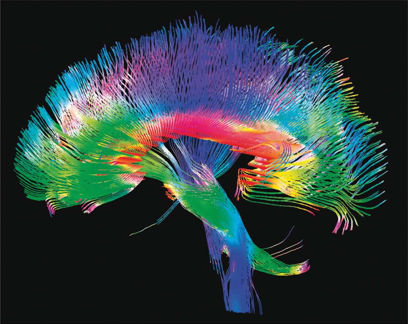
Genes help determine the nature of the brain's connectome, above, and the speed at which nerve transmissions can move along the connectome, between brain centres. Genes also influence brain plasticity via new stem cell production controlling the ability to form new memories.

Genes influence the ability to transfer attention to the more salient aspects of one's environment. This ability has always been a crucial survival trait.

Genes influence both the soundness of a person's feelings and intuition, and the trust that a person has in his own feelings. The sounder the intuition, the better able one is to predict future events and trends. Such prediction has always been a crucial survival trait.
Centre for Motivation? SD

An individual's strength and soundness of motivation helps to determine his ultimate destiny in life -- regardless of the underlying cultural substrate. Scientists are beginning to better understand the brain mechanisms of motivation.
Using images obtained from the MRI scans taken during the test, Mathias Pessiglione and his team identified a general motivational system in the depths of the brain, i.e. a structure capable of activating any effort type, both mental (concentrating on the task in hand) or physical (lifting a load). The researchers observed that the ventral striatum was activated in proportion to the amount of money involved: the higher the degree of motivation, the higher the activation level. Furthermore, the ventral striatum is connected to the median part of the striatum (the caudate nucleus) when the task to be performed is cognitively difficult (when the physical size and the numerical value of the numbers did not correspond). This ventral region solicits the lateral part of the striatum (the putamen) when the difficulty is motor-related (when the handle had to be squeezed very tightly).
The researchers suggest that the expectation of a reward is encoded in the ventral striatum, which can then drive either the motor or cognitive part of the striatum, depending on the task, in order to boost performance. _SD
More at PLoS Biology

Understanding the brain requires a certain level of comprehension of mechanisms operating at multiple levels simultaneously, from the molecular to the behavioural. It also requires an apprehension of circular causality, without which the brain could not function.

Each of us has a unique brain. The uniqueness of our brains is guaranteed by uniquely complex genetic sequences and mechanisms, but also by the unique environments within which these genetic mechanisms have played out.

We have barely begun to learn how our brains create us and our world. It is a great challenge, made all the greater by the incalculable variety that we comprise.

Labels: , , ,

Bookmark and Share

29 November 2011

Why Is it Important to Understand the Brain?

The human brain contains 100 billion (10^11) neurons, which combine to form almost 1 quadrillion (10^15) electro-chemical connections. Neurons are also affected by chemical signals that come via the blood, interstitial tissues, and glial cells. If we had to understand all the activity in the brain in order to understand the brain itself, we would be lost.

Fortunately, the brain organises itself in specific ways which simplify the task of discovering how the brain works.
2007 M. Raichle PNAS

The image above reveals particular nodes which participate in important brain networks. It is important that these nodes are able to communicate with other nodes participating in specific networks. Loss of nodes -- or the communication links between them -- can have devastating effects on normal brain function.
2011 van den Heuvel et Sporns Jnl Neurosci
The image above reveals the complexity of an average "connectome" which intervenes between the brain nodes participating in the 12 most important brain networks -- as measured by numbers of connections and activity levels. These networks begin to develop sometime between the 20th and 36th weeks of pregnancy.

Teasing out these connections, and following their activity in real time, is quite difficult work. But it is nothing when compared to the effort involved if one tried to follow the activity of 100 billion neurons simultaneously.

We can understand what happens in a normal human brain when the interconnections are disrupted, by looking at the brain under general anaesthesia.
Steven Laureys, who leads the Coma Science Group at the University of Liège in Wallonia, Belgium, looked at what happens during propofol anaesthesia when patients descend from wakefulness, through mild sedation, to the point at which they fail to respond to commands. He found that while small "islands" of the cortex lit up in response to external stimuli when people were unconscious, there was no spread of activity to other areas, as there was during wakefulness or mild sedation (Frontiers in Systems Neuroscience, vol 4, p 160). _NewScientist
So it is not only the ability of the brain nodes to function that counts, it is also vital that the nodes be able to communicate with each other. Depending upon which nodes or interconnections are disrupted, different types of alteration in normal brain function will take place.

This idea is crucial to understanding future modes of mass manipulation which will inevitably be utilised in the near future, by a wide range of groups with special and vested interest in the control of human populations. We know that it is possible to either inhibit or enhance the function of specific brain nodes using transcranial magnetic stimulatin (TMS) or transcranial DC stimulation. Understanding how the (temporary) loss of one specific node influences the function of the brain as a whole will give brain manipulators a wide range of approaches toward altering behaviour.

But there are far more powerful possibilities for influencing brain behaviour coming our way:
One scenario he imagines would make use of biological proteins manufactured with information-processing technology to deliver effects that could be triggered by electromagnetic stimulation. He imagined that they could be used in a club environment where the DJ would release nanoparticles that the audience could ingest. These could then be used to trigger the desired state at a particular point during his or her set using an electrical stimulus (from a headset) into the crowd's brains. _Wired
There is the idea of the nano-bio-info-cogno convergence, which opens the doors to mass manipulation of consciousness never possible before now.
The National Science Foundation (NSF) and a formidable-sounding government subcommittee called the National Science and Technology Council on Nanoscale Science, Engineering, and Technology have published a number of reports exploring the convergence of the NBIC technologies as the result of a series of conferences between 2001 and 2006. The chief application areas they’ve identified include:

• Expanding human cognition and communication,
• Improving human health and physical capabilities,
• Enhancing group and societal outcomes,
• Strengthening national security, and
• Unifying science and education.

The convergence, these reports suggest, will be based on the “unity of nature at the nanoscale” along with technology integration at the nanoscale, key transforming tools, and the pursuit of improvements in human performance. “A revolution is occurring in science and technology, based on the recently developed ability to measure, manipulate and organize matter on the nanoscale — 1 to 100 billionths of a meter,” writes William Sims Bainbridge, co-director of Human-Centered Computing at the NSF and co-editor with Mihail Roco of several NSF publications on NBIC. “At the nanoscale, physics, chemistry, biology, materials science, and engineering converge toward the same principles and tools. As a result, progress in nanoscience will have very far-reaching impact.” _H+Mag
Of course, when you read recommendations for "expanding human this," ... "improving human that," ... "enhancing human such," ... and so on, remember that when it is being done to you by powerful groups with vested interests, the more accurate word is "altering human this, that, and such." Presumably, the altering being done is to meet certain goals which you yourself did not necessarily formulate or put forth.

Powerful new tools of chemical synthesis, simultaneous brain imaging and manipulation, nano-drug delivery systems, and better cognitive understandings of how the brain works, all allow for powerfully convergent forms of manipulation which can only grow more powerful and specific over time.

Remember, though, that at the same time as the tools for group mind manipulation grow more powerful, the tools for self-understanding and self-control are also growing more powerful.

While legitimate uses for mind control may be set forth in national and international law -- to control episodes of deadly riots and insurrection, for example -- there is always the question of who is to watch the watchers? Even in the most benign and benevolent government, the human temptation to gain an advantage is always present. Wise governments are set up to make it very difficult for individuals and small groups of conspirators to gain control.

But have you seen any wise governments lately? Probably not. Which leaves protecting oneself from the coming tsunami of powerful group manipulation tools up to concerned individuals and groups who will probably not be government affiliated or supported.

We will return to this topic -- and ways to protect oneself in the face of these technological advances -- in the future.

Labels: , ,

Bookmark and Share

22 November 2011

Meditation as Treatment for Schizophrenia, Autism, Alzheimer's?

Dr. Judson Brewer, medical director of the Yale Therapeutic Neuroscience Clinic, and his colleagues asked 10 experienced meditators and 13 people with no meditation experience to practice three basic meditation techniques: concentration, loving-kindness, and choiceless awareness.

...In a report published today in the Proceedings of the National Academy of Sciences, Brewer and his team report that the experienced meditators had decreased activity in an area of the brain called the default mode network, a region that is usually at work when the mind wanders. Even when the meditators weren't meditating, this region of their brain was much quieter than in their inexperienced counterparts. _ABCNews
The areas shaded in blue highlight areas of decreased activity in the brains of meditators

The Yale team conducted functional magnetic resonance imaging scans on both experienced and novice meditators as they practiced three different meditation techniques.

They found that experienced meditators had decreased activity in areas of the brain called the default mode network, which has been implicated in lapses of attention and disorders such as anxiety, attention deficit and hyperactivity disorder, and even the buildup of beta amyloid plaques in Alzheimer's disease. The decrease in activity in this network, consisting of the medial prefrontal and posterior cingulate cortex, was seen in experienced meditators regardless of the type of meditation they were doing.

The scans also showed that when the default mode network was active, brain regions associated with self-monitoring and cognitive control were co-activated in experienced meditators but not novices. This may indicate that meditators are constantly monitoring and suppressing the emergence of "me" thoughts, or mind-wandering. In pathological forms, these states are associated with diseases such as autism and schizophrenia.

The meditators did this both during meditation, and also when just resting — not being told to do anything in particular. This may indicate that meditators have developed a "new" default mode in which there is more present-centered awareness, and less "self"-centered, say the researchers. _MedicalXpress
In a similar vein the University of Wisconsin is planning a study early next year to investigate the neurological effects of meditation and yoga with veterans.

It is thought mindfulness meditation holds promise for post-traumatic stress disorder (PTSD), which provokes intrusive thoughts, emotional numbness and hypervigilance.

Mindfulness-based cognitive therapy (MBCT), which combines meditation with orthodox 'thought training', is already recommended for depression in Britain and is available on the NHS. _DailyMail

Related research:
Fourteen meditation practitioners performed breath-focused meditation while undergoing fMRI scanning. When participants realized their mind had wandered, they pressed a button and returned their focus to the breath. The four intervals above were then constructed around these button presses. We hypothesized that periods of mind wandering would be associated with default mode activity, whereas cognitive processes engaged during awareness of mind wandering, shifting of attention and sustained attention would engage attentional subnetworks. Analyses revealed activity in brain regions associated with the default mode during mind wandering, and in salience network regions during awareness of mind wandering. Elements of the executive network were active during shifting and sustained attention. Furthermore, activations during these cognitive phases were modulated by lifetime meditation experience. These findings support and extend theories about cognitive correlates of distributed brain networks. _Abstract_Hasenkamp 2011 j. neuroimage Emory U.

Depression and the default mode network:
Major depressive disorder (MDD) has been characterized by excessive default-network activation and connectivity with the subgenual cingulate. These hyper-connectivities are often interpreted as reflecting rumination, where MDDs perseverate on negative, self-referential thoughts. However, the relationship between connectivity and rumination has not been established. Furthermore, previous research has not examined how connectivity with the subgenual cingulate differs when individuals are engaged in a task or not. The purpose of the present study was to examine connectivity of the default network specifically in the subgenual cingulate both on- and off-task, and to examine the relationship between connectivity and rumination. Analyses using a seed-based connectivity approach revealed that MDDs show more neural functional connectivity between the posterior-cingulate cortex and the subgenual-cingulate cortex than healthy individuals during rest periods, but not during task engagement. Importantly, these rest-period connectivities correlated with behavioral measures of rumination and brooding, but not reflection. _OxfordJournals

This is a lot of material to take in at once -- particularly if you are not familiar with the concept of the "default mode network." But understanding this concept can make a big difference in your life, and in those lives which you may influence along the way.

The default mode network is a "stand-by" brain network, which is active when you are not attending to anything. It is a state of the wandering mind, which all too often falls into repetitive thought patterns which are too often dysfunctional for many people.

The studies above reveal that meditation practise can change the circuits involved in the default mode network in a way that tends to reduce brooding, intrusive thoughts, and rumination -- even during times when one is not meditating. Self-monitoring and cognitive control during default mode activation was increased in meditators, although the overall intensity of default mode network function was decreased.

This is crucial: The idle mind may not be a quiet or relaxed mind. In fact, it is often a tortured or depressed mind, which over time can make chronic diseases of the brain and mind more likely to set in. If you want your mind to be relaxed when it falls into its inevitable periods of default mode, you may want to consider meditation training.

Labels: , , , ,

Bookmark and Share

18 November 2011

Genes Coming out of Nowhwere? Our Changing Brains

A few years ago scientists found that....once in awhile in the cells of all living things bits of once-quiet stretches of DNA sometimes spontaneously assemble themselves into genes. Such "de novo" genes may go on to play significant roles in the evolution of individual organisms—even humans.

...When an international team of researchers scanned the human genome for de novo genes, however, they putatively uncovered 60, three times more than once estimated. More surprising, many of these genes are active in the cerebral cortex, suggesting that de novo genes might have played a key role in the evolution of the human mind. _SciAm

We are learning more and more ways that our genes can change and vary -- changing who we are, and making us different from our ancestors and our fellow travelers. Evolution did not stop with the coming of civilisation -- it may have just gotten started.

Neurological variants such as autism and synaesthesia may be tentative "attempts" by evolution to create new species of human.
"If you think of ideas as being enshrined in neural populations in the brain, if you get greater cross-connectivity [in synaesthesia] you're going to create a propensity towards metaphorical thinking," he [Vilayanur Ramachandran] said. He suggested that this ability to link dissimilar concepts is what created a "huge explosion of abilities that characterise the human brain". _NewScientist
All it might take would be a basic change in the way the brain prunes its neurons in early development and in adolescence, to create a breed of human that thinks in significantly different ways. These changes might come about from the emergence of de novo genes, or via the modification or silencing of older, more ancient genes. But the end result might well be something quite remarkable.

Scientists are learning how to look at a genetic sequence and predict what the individual will be like, based upon those genes. Yes, we know that such a thing is actually far beyond the state of modern genetics and epigenetics, but suppose they can partially succeed at that goal. That would mean that an embryo's genes could be sampled in utero, and we would know if the coming child is likely to diverge from the "standard human genome." What would you do?

Imagine a new species of humans growing up at our feet -- all with at least the genius of a Mozart or an Einstein -- with the potential to revolutionise our world. For a short time they would be vulnerable to our wishes. But soon, they would be well beyond our reach.

It sounds like science fiction, but in many ways it is a far more likely scenario than the ideas of a superhuman machine intelligence that spawns "the singularity."

If modern humans were more homogeneous than they are, the possiblity of such an emerging, advanced, new human species would be low. But given the rather large differences that exist between different populations of modern humans already, it is almost easy to accept the idea of evolutionary divergence of human populations.

As humans gain a firmer grasp of the tools of genetics and epigenetics -- as well as environmental manipulations -- the possibility of an emerging altered subspecies of humans with particular niche advantages, grows stronger.

Labels: ,

Bookmark and Share

07 November 2011

To Be a Zombie, or Not to Be a Zombie: That is the Question

Default Mode Network Background

The "default mode network" of the brain is particularly active when the brain is resting, waiting for something "important" to do. The brain never goes completely idle, but instead goes into a stand-by mode. When we pay particular attention to things, the stand-by network goes idle to allow other parts of the brain to work. But what happens when the stand-by network is allowed to push the other brain networks around?.
A study carried out by a team at the Centre de Recherche en Neurosciences de Lyon (led by Tomas Ossandon and managed by Jean-Philippe Lachaux, Research Director at Inserm and Karim Jerbi, Research Leader at Inserm) has just revealed how this network interferes with our ability to pay attention, by assessing the activity of the human brain's default-mode network neurons on a millisecond scale for the first time ever, in collaboration with Philippe Kahane's epilepsy department in Grenoble.

The results unambiguously illustrate that whenever we look for an object in the area around us, the neurons of this default-mode network stop their activity. Yet, this interruption only lasts for the amount of time required to find the object: in less than a tenth of a second, after the object has been found, the default-mode network resumes its activity as before. And if our default-mode network is not sufficiently deactivated, then we will need more time to find the object. These results show that there is fierce competition for our attentional resources inside our brain which, when they are not used to actively analyse our sensorial environment, are instantaneously redirected towards more internal mental processes. _SD
Jnl Neuroscience Abstract
DMN [Default Mode Network] deactivation encodes the extent and efficiency of our engagement with the external world. Furthermore, our findings reveal a pivotal role for broadband gamma modulations in the interplay between task-positive and task-negative networks mediating efficient goal-directed behavior and facilitate our understanding of the relationship between electrophysiology and neuroimaging studies of intrinsic brain networks.

We are learning more about the perpetual tug-of-war that goes on between the networks of the brain. The brain is an insatiable consumer of the body's energy -- consuming 20% of O2 and glucose supplies. But if you actually think -- unlike most humans -- your brain will consume more. It is easier for the brain to do nothing, although even to do nothing the brain must still consume a lot of energy. The thinking networks of the brain require training and regular exercise, just as the muscles of the body require regular upkeep. If one does not continually train the cognitive networks, the default mode network will assume more influence.
Meta-Analysis of the default mode network Connectivity patterns

DocStoc Default Mode Network embed

The default mode is extremely important, in terms of saving energy and in terms of "resetting" or clearing the mind for whatever new situations may come up. At its best, the DMN opens the door to creative mental activity such as "lateral thinking." At its worst, when indulged too much, the DMN can make humans indistinguishable from zombies.

But when the situation calls for immediate planning or focus, the default mode may get in the way of your ability to achieve a clear conception of your situation or to formulate workable plans. Before that happens, one needs to make the choice whether to exercise the cognitive, planning, and decision-making networks of the brain, or to allow the default mode to occupy more and more of one's time. That choice is usually made very early in life, based upon a wide range of genetic and environmental factors.

For many things in our lives, we have no meaningful choice. That is why it is important to exercise choice when we can.

Labels: , , ,

Bookmark and Share

31 October 2011

Programmed to be Zombies? Stealth Genetics of Brain Transformation

Once thought to be rare, these events actually take place surprisingly often. According to one recent estimate, they occur in many or most brain cells, perhaps several hundred times within each cell...Many of the insertion sites were located within genes that play key roles in normal brain function. These include genes encoding receptors for the neurotransmitter dopamine and membrane transporters that mop up neurotransmitter molecules from the spaces between neurons after their signaling is complete... Others were found in genes encoding regulatory proteins that are linked to psychiatric illnesses such as schizophrenia and the developmental disorder Smith–Magenis syndrome. _SciAm
Slow Progressive Stealth Zombie Transformation

What if the people around us are not who we think they are? What if we continue to see them as someone they once resembled, while they have been subtly changing over time to something else?

Humans are defined by their brains. We know that their brains are shaped by a host of chance events involving genes, experiences, accidents etc. But what if these brains are changing in ways we do not understand, in ways we cannot control, so that over time these persons around us have changed into something else? I am not talking about diseases of the brain, but something much more subtle.

Here is a glimpse into the genetics of stealthy brain transformation which may help explain why that person next to you is not who you think they are:
Mobile DNA molecules that jump from one location in the genome to another may contribute to neurological diseases and could have subtle influences on normal brain function and behavior, according to a study published October 30 in Nature. (Scientific American is part of Nature Publishing Group.)

Retrotransposons are mobile genetic elements that use a copy-and-paste mechanism to insert extra copies of themselves throughout the genome. First discovered in plants about 60 years ago, they are now known to make up more than 40 percent of the entire human genome and may play an important role in genome evolution.

...The researchers also found that there was far more jumping-gene activity in the hippocampus than in the caudate nucleus. This is interesting, because the hippocampus is known to be critical for learning and memory, and is widely thought to be one of the few parts of the brain that continues to produce new cells throughout life. "It is tempting to speculate that genetic differences between individual neurons could impact memory," Faulkner says, "but we have no evidence yet that this is the case."

Retrotransposons are normally silenced to prevent harmful mutations from occurring in egg and sperm cells, but are mobilized during certain stages of brain development, when neurons are being produced from dividing stem cells. Retrotransposons then take the opportunity to jump at random into parts of the chromosome that have been opened up for DNA replication.

As well as generating mutations by inserting themselves into and disrupting genes, retrotransposons can alter gene activity if inserted into adjacent regulatory regions of DNA.

...Once thought to be rare, these events actually take place surprisingly often. According to one recent estimate, they occur in many or most brain cells, perhaps several hundred times within each cell
. Each neuron is likely subjected to a unique combination of insertions, leading to a genetic variability within populations of cells.

The full significance of this "genomic plasticity" is still not clear, but the authors suggest that it could influence brain development and behavior. It may, for example, partly account for the differences in brain structure and behavior between identical twins, and could even affect thought processes by subtly influencing the changes in nerve cell connections that occur with experience. _SciAm

It is becoming more and more difficult to claim that all humans are essentially the same, genetically. In fact, it is becoming more difficult to say that a person is the same today, genetically, as he was yesterday. Conventional methods of genomic sequencing do not detect many of these subtle genetic and epigenetic differences, suggesting the need for more sophisticated tools and screening methods.

One thing is certain: This changing nature of the brain -- the core of a human's self and being -- will require some rethinking of how societies should be organised. Humans with changing brains will need to be raised and taught to live in a dynamic and changing society. They must be given tools of competence, self reliance, and resilience. And they must be given the freedom to adapt to the inevitable changes which always occur.

Just the opposite of the traits one sees in the sheltered, academically lobotomised psychological neotenate -- who demands to be taken care of his entire life.

We can no longer afford this longing for dependency and stasis which defines most modern welfare state mentalities. This headlong rush for security, this raucous cry to be taken care of by an all-powerful state -- we cannot afford this delusional belief any longer. In a world of clashing cultures, this whining chorus of wankers is a suicidal lullaby. Particularly when the core populations of these affluent societies is shrinking so quickly.

Humans must learn to expect massive, dynamic changes over the course of their lives, both outside themselves and inside themselves. They must learn to be prepared to meet these changes on their own terms.

Retrotransposons and Human Genome Evolution

Abstract of Nature study:
Retrotransposons are mobile genetic elements that use a germline ‘copy-and-paste’ mechanism to spread throughout metazoan genomes1. At least 50 per cent of the human genome is derived from retrotransposons, with three active families (L1, Alu and SVA) associated with insertional mutagenesis and disease2, 3. Epigenetic and post-transcriptional suppression block retrotransposition in somatic cells4, 5, excluding early embryo development and some malignancies6, 7. Recent reports of L1 expression8, 9 and copy number variation10, 11 in the human brain suggest that L1 mobilization may also occur during later development. However, the corresponding integration sites have not been mapped. Here we apply a high-throughput method to identify numerous L1, Alu and SVA germline mutations, as well as 7,743 putative somatic L1 insertions, in the hippocampus and caudate nucleus of three individuals. Surprisingly, we also found 13,692 somatic Alu insertions and 1,350 SVA insertions. Our results demonstrate that retrotransposons mobilize to protein-coding genes differentially expressed and active in the brain. Thus, somatic genome mosaicism driven by retrotransposition may reshape the genetic circuitry that underpins normal and abnormal neurobiological processes.
More: Scientists key on changes in brain gene expression over the lifetime
In the studies, published in the Oct. 27 Nature, researchers focused not on DNA — virtually every cell’s raw genetic material is identical — but on when, where and for how long each gene is turned on over the course of a person’s life. To do this, the researchers measured levels of mRNA, a molecule whose appearance marks one of the first steps in executing the orders contained in a gene, in postmortem samples of donated brains that ranged in age from weeks after conception to old age.

...To see what those genes were up to, Šestan’s study examined mRNA levels of different genes in 57 brain samples. The team divided the brain tissue up by region, so they were also able to get an idea of genes’ behavior in different parts of the brain. A parallel study, headed by Joel Kleinman of the National Institute of Mental Health in Bethesda, looked at gene behavior in 269 brain samples from a single region called the prefrontal cortex that also spanned the lifetime.

This approach allowed the researchers to get access to the brain that had previously been impossible. _Sciencenews

We also need to remember that drug use triggers changes in brain gene expression -- some of these changes can be long-term or even "permanent." This is particularly tragic in the case of fetal brain exposure to drugs such as alcohol, methamphetamine, crack cocaine, etc. in the womb. But adolescents and young adults are also quite vulnerable to changes in brain gene expression from drug use. Bonus question: What are the most effective cultures and incubators for drug use in societies? Schools, and anywhere young peers are concentrated and on their own.

Long term behavioural change following ingestion of magic mushrooms More

Brain changes from cannabis use

And there are always new party drugs coming down the pipeline which will have unknown short and long-term effects on the brain, and brain gene expression.

Some people are naturally programmed to be zombies. Other people have to work at it.

Labels: , ,

Bookmark and Share
Older Posts
Al Fin Main Page
Enter your Email


Powered by FeedBlitz
Google
WWW AL FIN

Powered by
Blogger

``