In Search of Dr. McCoy's Scanner Wand

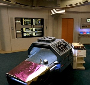
Medical imaging in the Star Trek world was far advanced over anything available today. Even so, there are some amazing developments in medical imaging that promise even better technologies in the near future.
This Newswise newsrelease reports on advanced software developed at BYU in Utah, to allow physicians to "call up" 3-dimensional images of any part of a patient's anatomy, from a CT scan, MRI scan, or similar data:
The tool, dubbed “Live Surface” by its creators BYU professor William Barrett and graduate student Chris Armstrong, also has special-effects applications – in similar fashion it can be used to extract a single actor’s performance or inanimate objects from video clips.Source.
“The main goal in developing Live Surface was to give the physician a powerful, practical tool that can be used interactively,” said Barrett, explaining that existing software and techniques that are used to give doctors a look at a patient’s anatomy are either too simplistic or take too long to be of immediate use. “A program like this has to be incredibly fast and very interactive, or else it’s very frustrating for the user, who currently has to go get a sandwich and come back before he has what he wants.”
Live Surface has the additional benefit of allowing users to easily isolate “tricky” anatomy such as soft tissue – blood vessels, hearts and muscles – that a lot of other techniques can’t readily extract, said Barrett.
“The hard stuff (meaning bones) is easy to see using traditional methods, but even there, the simpler techniques sometimes overestimate, underestimate, or fuse joints, whereas Live Surface neatly and accurately separates them,” said Barrett. “Our program also provides more robust isolation of soft tissue, which is quite a breakthrough.”
The BYU software works by extracting information from data collected in 3-D volumes – CT scans, MRIs or 3-D ultrasounds. With a click and drag of the mouse, a user identifies the object he wishes to extract. Next, he identifies those portions of the data that surround the object. Immediately, the desired object is extracted from the data.
“This is the object I want, and this is not the object I want. And in less than half a second, it pulls the object you want out of the data,” said Armstrong, who will present the computer science research behind Live Surface at the International Workshop on Volume Graphics 31 in Boston. “It’s really that simple.”
....After a surgeon had extracted a 3-D image of a person’s heart or brain, for example, the image could then be projected onto the patient’s body, fitted to create a road map for the surgeon as he operated. Additionally, doctors could use the tool to make better diagnoses after visualizing a patient’s organs from multiple angles or do a better job of locating cancerous tumors.
Research for the software was partially funded by Adobe, makers of the popular image-editing program Photoshop. Barrett’s lab has had a long-running relationship with Adobe – Live Surface builds on Barrett’s development of Intelligent Scissors, a program that allows users to quickly pull 2-D objects out of images. The program, renamed “magnetic lasso,” was incorporated into 5.0 and subsequent versions of Photoshop and is used by millions of designers, artists and photographers.
It is easy to see how this technology would allow a surgeon to "practice" a procedure on a specific individual, using virtual surgery techniques combined with a CT scan or MRI scan of the involved part of the body. Difficult procedures could be stored in software libraries for training surgical residents.
Similar technology is being used to perform virtual colonoscopies, to substitute for the lengthy and uncomfortable screening procedure of colonoscopy using an endoscope. Eventually, a whole body scan might become quick enough, easy enough, and inexpensive enough to provide regular and reliable medical screening for individuals whose genetic tests indicate they are at increased risk.
Labels: medical imaging, star trek science

