06 November 2012

Brain Networks in Anesthesia, Sleep, and Waking

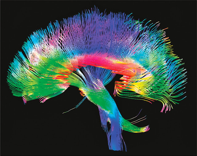
Recent research collaboration between MIT, MGH, and Harvard Medical School has substantiated earlier findings on the mechanism of loss of consciousness in the human brain during general anesthesia.

Using sophisticated research tools including EEG and fMRI, they found that as consciousness was lost, different parts of the brain became "disconnected" from each other, even though particular areas of the brain continued to function.
...the time when consciousness was lost did coincide with a significant change in the overall structure of brain activity. While electrical activity in the conscious brain appears to be disorganized with no apparent regular patterns, at the point when study participants lost consciousness, their brain activity began to show regular oscillations between states of activation and deactivation.

..."These deactivated or silent periods of brain activity occur at different times in different brain regions, so communication between regions is interrupted" says Laura Lewis, co-lead author of the report. "It's as if one brain region is in Boston and the other is in Singapore – they can't make phone calls to each other because one is asleep when the other is awake." While this slow oscillation pattern has been previously observed in humans who are asleep or under ansethesia, this is the first study to record neuronal activity during the transition to unconsciousness, so it is the first to match the onset of this pattern with the loss of consciousness, she adds. _MXP

Quick synopsis of study:
The team asked each patient to respond to a sound as they drifted off. At the moment they stopped responding, Lewis and Purdon saw a dramatic change in neuronal activity across the cortex. Slow wave oscillations – the brainwaves that occur in deep, non-dreaming sleep – grew almost immediately.

Locally, these slow waves were in sync and neurons near each other coordinated their activity to correspond with the peaks and troughs of the waves they encountered, meaning continued communication. However, the slow waves were not in sync across the entire cortex.

While conscious, different regions of the cortex fire at the same time, so neurons can communicate over long distances if necessary... out-of sync slow waves make long distance communication near impossible. _NewScientist


PNAS study abstract for above research

More reading on brain networks in general anesthesia:

The Mystery Behind Anesthesia -- an article published almost a year ago in Technology Review, describing earlier research by the same team

Going Under -- a nice article from Science News published about 18 months ago, looking at earlier research from the above MIT/MGH/Harvard team and other competing research teams from the UK and U. Michigan / Cornell.

One cannot help considering the parallels between general anesthesia and normal sleep. Here are two articles that look at brain network activity in relation to sleep and consciousness:

The Process of Awakening

Development of Large Scale Functional Brain Network During Human Non-REM Sleep -- looks at activity of brain networks around the time of onset of sleep

You may ask why one should bother trying to understand how brain networks are put under, fall asleep, or wake up -- and how all that relates to normal brain network function during consciousness. The answer to that question comes along with all the things we will be able to do, once we understand.

It might be easier to understand how the human brain works if we had more sophisticated analytical and synthetic tools than the human brain to work with. As it is, we will have to muddle along trying out different theories until something "clicks into place." At that point, we should be able to design some pretty amazing brain assistants and brain substitutes.

Labels: ,

Bookmark and Share

22 October 2012

Brain Network Dynamics: Brain as Anti-Algorithm

Cognitive scientists are uncovering more secrets of the brain every day. One fascinating line of brain research involves how the brain forms categories and metaphors.
At the IMP in Vienna, neurobiologist Simon Rumpel and his post-doc Brice Bathellier have been able to show that certain properties of neuronal networks in the brain are responsible for the formation of categories. In experiments with mice, the researchers produced an array of sounds and monitored the activity of nerve cell-clusters in the auditory cortex. They found that groups of 50 to 100 neurons displayed only a limited number of different activity-patterns in response to the different sounds.

The scientists then selected two basis sounds that produced different response patterns and constructed linear mixtures from them. When the mixture ratio was varied continuously, the answer was not a continuous change in the activity patters of the nerve cells, but rather an abrupt transition. Such dynamic behavior is reminiscent of the behavior of artificial attractor-networks that have been suggested by computer scientists as a solution to the categorization problem. _SD

Here is the study abstract from Neuron:
The ability to group stimuli into perceptual categories is essential for efficient interaction with the environment. Discrete dynamics that emerge in brain networks are believed to be the neuronal correlate of category formation. Observations of such dynamics have recently been made; however, it is still unresolved if they actually match perceptual categories. Using in vivo two-photon calcium imaging in the auditory cortex of mice, we show that local network activity evoked by sounds is constrained to few response modes. Transitions between response modes are characterized by an abrupt switch, indicating attractor-like, discrete dynamics. Moreover, we show that local cortical responses quantitatively predict discrimination performance and spontaneous categorization of sounds in behaving mice. Our results therefore demonstrate that local nonlinear dynamics in the auditory cortex generate spontaneous sound categories which can be selected for behavioral or perceptual decisions. _Neuron Article Abstract
Here is a broader look at brain network dynamics in the context of decision making:

Cortical network dynamics of perceptual decision-making in the human brain

Brain cells work together in groups, in a dynamic fashion.

Spontaneous rhythmical activity occurs in groups of neurons -- whether artificially cultured in the lab, or in self-selected groups within a living brain.

When separated groups of neurons communicate with each other over a distance in the brain, they utilise a method of synchronous oscillations -- a language that scientists have just begun to understand.

Billions of dollars are spent every year on the quest to achieve human level artificial intelligence. Most of this research is based upon algorithmic design, utilising digital computers. But as anyone can see from looking over recent findings in the neuroscience of cognition, the brain is more of an anti-algorithm. The logic of brain network dynamics has almost nothing in common, conceptually, with the algorithmic basis of digital computing.

AI researchers have attempted to narrow the conceptual gap by utilising "neural net computing," "fuzzy logic computing," and "genetic algorithmic computing," to name three alternative approaches. And these alternative approaches are likely to be very helpful in both applied and theoretical computing and information science. But do they get AI researchers closer to the goal of human-level machine intelligence?

Probably not. Not even the startling potential of memristors and similar semiconductor devices are likely to close that gap appreciably.

As discussed recently in an article quoting quantum physicist David Deutsch, artificial intelligence research is desperately in need of better supporting philosophical structures.

Until then, it is likely that artificial intelligence research will continue to spin its wheels pursuing better algorithms to emulate the brain, without a good understanding of what the brain does.

It is possible to emulate the human brain, using an approach that depends to a limited extent upon algorithmic control, in conjunction with other conceptual methods. But not before researchers learn to approach the problem in entirely new ways, on new logical levels..

Introduction to brain oscillations video

Labels: , ,

Bookmark and Share

13 June 2012

Learn to Control Your Own Brain Emotion Networks w/ Real Time fMRI Neurofeedback

Neuro-researchers in the UK and the Netherlands helped establish real-time fMRI neurofeedback as a practical tool for self-controlling emotional networks in healthy subjects, back in 2009 (PDF). Since then, they have studied the use of real time fMRI feedback in the treatment of depression, with some positive results. Here is more from their most recent study:
a) Activation of the insular cortex (INS) bilaterally and the right ventral striatum (VS) supported the neurofeedback task, whereas the temporoparietal junctions (TPJ) of both hemispheres were deactivated. The TPJ is recognised as part of the brain’s “default mode network” that is deactivated during effortful tasks.
plosone

Depression is the mental disorder with the largest impact on public health. Up to 20% of the population suffers from a depressive episode at some point in their lives [1], and major depressive disorder (MDD) is a main source of disability for adults of working age in industrialized countries. At least 30% of patients with MDD do not respond to standard pharmacological and/or psychological treatments [2], and a considerable number of those who do respond initially go on to develop a chronic relapsing-remitting disorder. These patients with no or only a partial response to standard treatments often enter a vicious circle of psychosocial decline with further deterioration of their mood and level of functioning. To prevent relapses new therapeutic strategies have to be developed that aid the restructuring of cognitive schemas and might even prevent the formation and crystallization of dysfunctional thought patterns during early phases of depression.

...In the present study we localized areas responsive to positively valenced visual stimuli adapted from the International Affective Pictures System (IAPS) [15], [10] and then trained patients with unipolar depression to upregulate the activity in this target region over four sessions. We hypothesized that the combination of the physiological upregulation and the reinforced training of positive thought patterns would lead to an improvement of mood, which would not be seen in a control group that engaged in an emotion regulation protocol without neurofeedback. _PLoS ONE
Higher activation of right insula (INS), ventral striatum (VS), anterior cingulate cortex (ACC) and ventromedial prefrontal cortex (VMPFC) during presentation of positive compared to neutral images in the localiser runs (for full list of areas see Table 2). The localiser runs were effective in identifying brain areas responsive to positive images, which were used as target regions of interests (ROIs) for the subsequent neurofeedback procedure.
plosone

Patients in the NF group were trained to upregulate brain areas responsive to positive emotions using a procedure modeled on our previous work with healthy participants [10]. A target area was identified by the contrast between responses to positive and neutral images in a localizer scan to ensure that an area involved in positive emotion processing was selected. In the localizer scan, we assessed brain responses to positive, negative and neutral pictures by presenting four pictures of the same emotion category in blocks of 6 s (1.5 s per picture), alternating with a fixation baseline of 12 s. We presented 12 blocks per category in pseudorandom order. We used pictures from the IAPS [15] with negative (mean normative ratings for valence 2.8 [SD.42], arousal 5.63 [SD.55]), positive (valence 6.90 [.55], arousal 6.00 [.74]) and neutral valence (valence 5.45 [.56], arousal 3.44 [.47]). Pictures showed, for example, scenes of danger or disgust in the negative category, and scenes of romance including mild erotica or exciting sports in the positive category. After the localizer scan, patients were trained to upregulate the target area during three neurofeedback scans lasting ca. 7 minutes each per session (Fig. 1). Patients were informed about the general function of the target area but were not given any specific instructions about strategy. The task we set for them was to increase activity in the target area by as much and as consistently as possible.

...For the neurofeedback, a continuous signal from the target area (updated every TR and thus every 2 seconds) was displayed using the picture of a thermometer whose dial indicated the amplitude of the fMRI signal in the target area. Changes in the amplitude were indicated as the percent of signal change, calculated using the current signal intensity value and comparing it with the average value determined from the rest period immediately preceding each upregulation block. The scaling of the thermometer was in steps of 0.05%, with a maximum value of 0.5% (see Fig. 1). A change of background colour every 20 s indicated to participants whether their task was to regulate (green background) or rest (yellow background). The online GLM was computed with one predictor for the regulation state, convolved with a haemodynamic reference function. The top one-third (defined by the t value for the contrast between the regulation predictor and baseline) of the voxels from the target region was used to compute the feedback signal. For runs in which participants failed to upregulate the target area during the regulation periods (negative percent signal change), another target area was selected for the next run, using the cluster with the strongest activation for the regulation predictor. This adjustment in the target area was necessary in 15/32 (47.9%) of the sessions after the first NF run, and in 4 sessions after the second run. The reasons for this approach were two-fold. First, the adjustment of ROIs aided the shaping of mental strategies in the desired direction. Shaping is a common concept in the operant learning of a highly demanding task [11]. Secondly, our focus was not so much on the ability of participants to learn to regulate a specific brain region but on the effects of the NF training procedure on participants’ mood.

...Patients in the NF group reported initially using imagery of the positive scenes in the localizer scan in an attempt to increase activation in the target brain areas, but they later changed to evoking memories and imagery of autobiographically relevant material. For example, the happy memories that they reported as successful strategies included holidays, thoughts about their family being happy, and imagery of beautiful scenes from nature. Some patients attained good self-regulation of the target areas through mental simulation of future successes, and one patient successfully used imagery of an out-of-body experience. Conversely, during rest periods, the patients reported trying to “empty their thoughts” and to meditate. Patients in the IM group were instructed to engage in similar strategies as those reported by the NF patients. At debriefing, they confirmed that they had used these strategies. No patient reported any distress arising from the procedure.

...In the present study, four sessions of non-invasive fMRI-neurofeedback reduced the symptoms of depression with an effect size similar to those obtained with deep brain stimulation (DBS) [3]. Although the mental strategies of positive thoughts, memories, and imagery may have played a considerable part in this improvement, the neurofeedback procedure was crucial as evidenced by the absence of any clinical improvement in the control group. _PLoS ONE
More study details at the link above.

The researchers intend to expand their research in future studies so as to deal with possible confounders, and to introduce increased rigour via randomisation and more sophisticated control procedures.

There are many advantages in the ability to detect and shift the activity in one's own emotion networks. This could be true both in one's professional and personal life. It is also likely that the ability to control specific brain networks will facilitate learning -- particularly in difficult subjects where frustration and apprehension can be a factor.

fMRI neurofeedback is a bit unwieldy, given the bulk and expense of fMRI scanners. Sophisticated EEG neurofeedback can achieve essentially the same results with less expense, although the increased neuroscientific rigour of visualising fMRI network activation and deactivation, is an advantage in the research setting.

Once protocols are developed with fMRI, then parallel protocols using advanced -- but portable -- EEG can be cross-validated using data from the fMRI.

The microprocessor revolution has helped to shrink the size and reduce the weight of a wide range of sophisticated electronic devices. Home EEG equipment is already available which can be used in conjunction with a pad computer or a smart phone. As these devices improve -- and as the protocols for self-control of brain networks continue to be developed and improved -- expect to be able to teach your brain to control itself in more ways than you might currently imagine.

Labels: , ,

Bookmark and Share

30 December 2011

Fine Tuning Brain Ensembles with Glial Overseers

Glia cells, named for the Greek word for "glue," hold the brain's neurons together and protect the cells that determine our thoughts and behaviors, but scientists have long puzzled over their prominence in the activities of the brain dedicated to learning and memory. Now Tel Aviv University researchers say that glia cells are central to the brain's plasticity — how the brain adapts, learns, and stores information.

...Almost all neurodegenerative diseases are glia-related pathologies, Prof. Ben-Jacob notes. In epileptic seizures, for example, the neurons' activity at one brain location propagates and overtakes the normal activity at other locations. This can happen when the glia cells fail to properly regulate synaptic transmission. Alternatively, when brain activity is low, glia cells boost transmissions of information, keeping the connections between neurons "alive." _aftau.org

To understand the brain, one must conceptualise multiple levels of activity occurring simultaneously. The level where glial cells influence neurons and synaptic output is a relatively low to mid-level, but a crucial one. Neuroscience has barely begun to understand this basic interaction between cell types, and is forced -- as in this study -- to use neurocomputational models in an attempt to expand the comprehension of this complex give and take.
The brain is constituted of two main types of cells: neurons and glia. Neurons fire off signals that dictate how we think and behave, using synapses to pass along the message from one neuron to another, explains De Pittà. Scientists theorize that memory and learning are dictated by synaptic activity because they are "plastic," with the ability to adapt to different stimuli.

But Ben-Jacob and colleagues suspected that glia cells were even more central to how the brain works. Glia cells are abundant in the brain's hippocampus and the cortex, the two parts of the brain that have the most control over the brain's ability to process information, learn and memorize. In fact, for every neuron cell, there are two to five glia cells. Taking into account previous experimental data, the researchers were able to build a model that could resolve the puzzle.

The brain is like a social network, says Prof. Ben-Jacob. Messages may originate with the neurons, which use the synapses as their delivery system, but the glia serve as an overall moderator, regulating which messages are sent on and when. These cells can either prompt the transfer of information, or slow activity if the synapses are becoming overactive. This makes the glia cells the guardians of our learning and memory processes, he notes, orchestrating the transmission of information for optimal brain function. _aftau.org

PLoS CompBio
Looking at how synaptic connections change during learning and memory is a little bit like trying to understand what goes on inside an office building by studying a set of architectural blueprints and reviewing the utility bills. It provides a bare beginning, but it's a start.
Synaptic plasticity is the capacity of a preexisting connection between two neurons to change in strength as a function of neuronal activity. Because it admittedly underlies learning and memory, the elucidation of its constituting mechanisms is of crucial importance in many aspects of normal and pathological brain function. Short-term presynaptic plasticity refers to changes occurring over short time scales (milliseconds to seconds) that are mediated by frequency-dependent modifications of the amount of neurotransmitter released by presynaptic stimulation. Recent experiments have reported that glial cells, especially hippocampal astrocytes, can modulate short-term plasticity, but the mechanism of such modulation is poorly understood. Here, we explore a plausible form of modulation of short-term plasticity by astrocytes using a biophysically realistic computational model. Our analysis indicates that astrocytes could simultaneously affect synaptic release in two ways. First, they either decrease or increase the overall synaptic release of neurotransmitter. Second, for stimuli that are delivered as pairs within short intervals, they systematically increase or decrease the synaptic response to the second one. Hence, our model suggests that astrocytes could transiently trigger switches between paired-pulse depression and facilitation. This property explains several challenging experimental observations and has a deep impact on our understanding of synaptic information transfer. _PLoSCompBio

PLoS Comp Bio
By understanding how synaptic frequencies can be modulated by changes in the glial::neuronal interaction, scientists can begin to bridge upward to the next higher levels of cognition -- local and distant neuronal ensembles. No one said it would be easy.

Neuroscientists are forced to work with a variety of animal models, human brain imaging techniques, cell culture approaches, and computational models, in order to enlarge their understanding of the foundations of an incredibly complex, multi-layered, and time-dependent phenomenon -- consciousness. And at that, we are talking about the consciousness of lower animals.

PLoS article in full
Bonus link: Wim de Neys at U. Toulouse is attempting to clarify some of the insights that come from the work of Nobel Prize winner Daniel Kahneman. de Neys seems to want to place human intuition on a higher level than the one on which Kahneman's work suggests it belongs. Certainly humans are dependent upon their intuitions for most of the things they do and virtually all the choices they make. In the opinion of Al Fin cognitivists, such psychological research still suffers from the lack of a firm neuroscientific foundation.

Labels: , ,

Bookmark and Share

23 December 2011

Syncopated Ghost Whispers Haunt Your Internal Web

Long Distance Brain Network Macaque PNAS

Above, you see an early depiction of the "long distance network" of the brain, connecting different brain centres with each other. The complex visualisation was compiled using information obtained from the study of the macaque brain.
The Human Connectome Project is hard at work producing images such as this, using an MRI technique known as diffusion tensor imaging.
With 100 billion neurons, each with around 10,000 connections, mapping the human brain will be no easy feat, and charting every single connection could take decades. The HCP will tackle the lowest hanging fruit first: charting the major highways between different brain regions, and showing how these connections vary between individuals. To do this they will combine several imaging tools including something called diffusion MRI, which maps the structure of the white matter that insulates the "wires" of the brain, and also resting-state MRI, which measures how brain regions oscillate in unison as a result of shared connections. _NewScientist

Cortical parcellations (PDF) such as the above, use another MRI technique. This method of brain visualisation separates different cortical domains which serve particular functions.
These brain images are presented for purposes of orientation and grounding. They may help to picture the various nodes and connections presented in the abstracted images and schematics.
Above, you see some of the brain areas involved in three important brain networks: Default Mode, Salience, and Central Executive. When viewing such fMRI "activation" images, it is helpful to mentally superimpose the connections between the activated brain centres. More on the three pictured networks:
The default mode (DMN) or default brain network (shown in blue) is what your brain does when not engaged in specific tasks. It is the busy or active part of your brain when you are mentally passive. According to Bresslor and Brennon the “DMN is seen to collectively comprise an integrated system for autobiographical, self-monitoring and social cognitive functions.” It has also been characterized as responsible for REST (rapid episodic spontaneous thinking). In other words, this is the spontaneous mind wandering and internal self-talk and thinking we engage in when not working on a specific task or, when completing a task that is so automatized (e.g., driving a car) that our mind starts to wander and generate spontaneous thoughts.

...The salience network (shown in yellow) is a controllor or network switcher. It monitors information from within (internal input) and from the external world arounding us, which is constantly bombarding us with information. Think of the salience network as the air traffic controllor of the brain. Its job is to scan all information bombarding us from the outside world and also that from within our own brains. This controller decides which information is most urgent, task relevant, and which should receive priority in the que of sending brain signals to areas of the brain for processing.

...Finally, the central-executive network (CEN; shown in red) “is engaged in higher-order cognitive and attentional control.” In other words, when you must engage your concious brain to work on a problem, place information in your working memory as you think, focus your attention on a task or problem, etc., you are “thinking” and must focus your controlled attention. _BrainClockBlog
We have talked about the default mode network previously, and will devote future time to the integration of various overlapping -- as well as mutually exclusive -- networks.
Now, we are getting close to the "brass tacks" of how separate brain nodes communicate synchronously with each other via the connectome. The brain functions as a hierarchical network, and depends upon analogous -- but different -- mechanisms of ensemble activity at different levels of the hierarchy.
... when multiple neurons spread all over the brain are tuned in to a specific pattern of electrical activity at a specific frequency, then whenever that global activity pattern occurs, those neurons can act as a coordinated assembly."
The researchers pointed out that this mechanism of cell assembly formation via oscillatory phase coupling is selective. Two neurons that are sensitive to different frequencies or to different spatial coupling patterns will exhibit independent activity, no matter how close they are spatially, and will not be part of the same assembly. Conversely, two neurons that prefer a similar pattern of coupling will exhibit similar spiking activity over time, even if they are widely separated or in different brain areas. _SD
One of the many things that makes understanding the brain so difficult, is the fact that so many things are happening all at once, on so many different levels -- both in serial and parallel format. Almost all of the things that go on in the brain occur on the unconscious or subconscious levels. Consciousness, as we know it, is something of an over-rated evolutionary accident.

Video via Kevin at Brain Clock Blog

Finally, watch ghostly whispers moving through the human brain as it is put through its paces.

More information on brain networks at The Brain Clock Blog: The brain as a set of networks: Fine tunning your networks

Labels: , , ,

Bookmark and Share

29 November 2011

Why Is it Important to Understand the Brain?

The human brain contains 100 billion (10^11) neurons, which combine to form almost 1 quadrillion (10^15) electro-chemical connections. Neurons are also affected by chemical signals that come via the blood, interstitial tissues, and glial cells. If we had to understand all the activity in the brain in order to understand the brain itself, we would be lost.

Fortunately, the brain organises itself in specific ways which simplify the task of discovering how the brain works.
2007 M. Raichle PNAS

The image above reveals particular nodes which participate in important brain networks. It is important that these nodes are able to communicate with other nodes participating in specific networks. Loss of nodes -- or the communication links between them -- can have devastating effects on normal brain function.
2011 van den Heuvel et Sporns Jnl Neurosci
The image above reveals the complexity of an average "connectome" which intervenes between the brain nodes participating in the 12 most important brain networks -- as measured by numbers of connections and activity levels. These networks begin to develop sometime between the 20th and 36th weeks of pregnancy.

Teasing out these connections, and following their activity in real time, is quite difficult work. But it is nothing when compared to the effort involved if one tried to follow the activity of 100 billion neurons simultaneously.

We can understand what happens in a normal human brain when the interconnections are disrupted, by looking at the brain under general anaesthesia.
Steven Laureys, who leads the Coma Science Group at the University of Liège in Wallonia, Belgium, looked at what happens during propofol anaesthesia when patients descend from wakefulness, through mild sedation, to the point at which they fail to respond to commands. He found that while small "islands" of the cortex lit up in response to external stimuli when people were unconscious, there was no spread of activity to other areas, as there was during wakefulness or mild sedation (Frontiers in Systems Neuroscience, vol 4, p 160). _NewScientist
So it is not only the ability of the brain nodes to function that counts, it is also vital that the nodes be able to communicate with each other. Depending upon which nodes or interconnections are disrupted, different types of alteration in normal brain function will take place.

This idea is crucial to understanding future modes of mass manipulation which will inevitably be utilised in the near future, by a wide range of groups with special and vested interest in the control of human populations. We know that it is possible to either inhibit or enhance the function of specific brain nodes using transcranial magnetic stimulatin (TMS) or transcranial DC stimulation. Understanding how the (temporary) loss of one specific node influences the function of the brain as a whole will give brain manipulators a wide range of approaches toward altering behaviour.

But there are far more powerful possibilities for influencing brain behaviour coming our way:
One scenario he imagines would make use of biological proteins manufactured with information-processing technology to deliver effects that could be triggered by electromagnetic stimulation. He imagined that they could be used in a club environment where the DJ would release nanoparticles that the audience could ingest. These could then be used to trigger the desired state at a particular point during his or her set using an electrical stimulus (from a headset) into the crowd's brains. _Wired
There is the idea of the nano-bio-info-cogno convergence, which opens the doors to mass manipulation of consciousness never possible before now.
The National Science Foundation (NSF) and a formidable-sounding government subcommittee called the National Science and Technology Council on Nanoscale Science, Engineering, and Technology have published a number of reports exploring the convergence of the NBIC technologies as the result of a series of conferences between 2001 and 2006. The chief application areas they’ve identified include:

• Expanding human cognition and communication,
• Improving human health and physical capabilities,
• Enhancing group and societal outcomes,
• Strengthening national security, and
• Unifying science and education.

The convergence, these reports suggest, will be based on the “unity of nature at the nanoscale” along with technology integration at the nanoscale, key transforming tools, and the pursuit of improvements in human performance. “A revolution is occurring in science and technology, based on the recently developed ability to measure, manipulate and organize matter on the nanoscale — 1 to 100 billionths of a meter,” writes William Sims Bainbridge, co-director of Human-Centered Computing at the NSF and co-editor with Mihail Roco of several NSF publications on NBIC. “At the nanoscale, physics, chemistry, biology, materials science, and engineering converge toward the same principles and tools. As a result, progress in nanoscience will have very far-reaching impact.” _H+Mag
Of course, when you read recommendations for "expanding human this," ... "improving human that," ... "enhancing human such," ... and so on, remember that when it is being done to you by powerful groups with vested interests, the more accurate word is "altering human this, that, and such." Presumably, the altering being done is to meet certain goals which you yourself did not necessarily formulate or put forth.

Powerful new tools of chemical synthesis, simultaneous brain imaging and manipulation, nano-drug delivery systems, and better cognitive understandings of how the brain works, all allow for powerfully convergent forms of manipulation which can only grow more powerful and specific over time.

Remember, though, that at the same time as the tools for group mind manipulation grow more powerful, the tools for self-understanding and self-control are also growing more powerful.

While legitimate uses for mind control may be set forth in national and international law -- to control episodes of deadly riots and insurrection, for example -- there is always the question of who is to watch the watchers? Even in the most benign and benevolent government, the human temptation to gain an advantage is always present. Wise governments are set up to make it very difficult for individuals and small groups of conspirators to gain control.

But have you seen any wise governments lately? Probably not. Which leaves protecting oneself from the coming tsunami of powerful group manipulation tools up to concerned individuals and groups who will probably not be government affiliated or supported.

We will return to this topic -- and ways to protect oneself in the face of these technological advances -- in the future.

Labels: , ,

Bookmark and Share

22 November 2011

Meditation as Treatment for Schizophrenia, Autism, Alzheimer's?

Dr. Judson Brewer, medical director of the Yale Therapeutic Neuroscience Clinic, and his colleagues asked 10 experienced meditators and 13 people with no meditation experience to practice three basic meditation techniques: concentration, loving-kindness, and choiceless awareness.

...In a report published today in the Proceedings of the National Academy of Sciences, Brewer and his team report that the experienced meditators had decreased activity in an area of the brain called the default mode network, a region that is usually at work when the mind wanders. Even when the meditators weren't meditating, this region of their brain was much quieter than in their inexperienced counterparts. _ABCNews
The areas shaded in blue highlight areas of decreased activity in the brains of meditators

The Yale team conducted functional magnetic resonance imaging scans on both experienced and novice meditators as they practiced three different meditation techniques.

They found that experienced meditators had decreased activity in areas of the brain called the default mode network, which has been implicated in lapses of attention and disorders such as anxiety, attention deficit and hyperactivity disorder, and even the buildup of beta amyloid plaques in Alzheimer's disease. The decrease in activity in this network, consisting of the medial prefrontal and posterior cingulate cortex, was seen in experienced meditators regardless of the type of meditation they were doing.

The scans also showed that when the default mode network was active, brain regions associated with self-monitoring and cognitive control were co-activated in experienced meditators but not novices. This may indicate that meditators are constantly monitoring and suppressing the emergence of "me" thoughts, or mind-wandering. In pathological forms, these states are associated with diseases such as autism and schizophrenia.

The meditators did this both during meditation, and also when just resting — not being told to do anything in particular. This may indicate that meditators have developed a "new" default mode in which there is more present-centered awareness, and less "self"-centered, say the researchers. _MedicalXpress
In a similar vein the University of Wisconsin is planning a study early next year to investigate the neurological effects of meditation and yoga with veterans.

It is thought mindfulness meditation holds promise for post-traumatic stress disorder (PTSD), which provokes intrusive thoughts, emotional numbness and hypervigilance.

Mindfulness-based cognitive therapy (MBCT), which combines meditation with orthodox 'thought training', is already recommended for depression in Britain and is available on the NHS. _DailyMail

Related research:
Fourteen meditation practitioners performed breath-focused meditation while undergoing fMRI scanning. When participants realized their mind had wandered, they pressed a button and returned their focus to the breath. The four intervals above were then constructed around these button presses. We hypothesized that periods of mind wandering would be associated with default mode activity, whereas cognitive processes engaged during awareness of mind wandering, shifting of attention and sustained attention would engage attentional subnetworks. Analyses revealed activity in brain regions associated with the default mode during mind wandering, and in salience network regions during awareness of mind wandering. Elements of the executive network were active during shifting and sustained attention. Furthermore, activations during these cognitive phases were modulated by lifetime meditation experience. These findings support and extend theories about cognitive correlates of distributed brain networks. _Abstract_Hasenkamp 2011 j. neuroimage Emory U.

Depression and the default mode network:
Major depressive disorder (MDD) has been characterized by excessive default-network activation and connectivity with the subgenual cingulate. These hyper-connectivities are often interpreted as reflecting rumination, where MDDs perseverate on negative, self-referential thoughts. However, the relationship between connectivity and rumination has not been established. Furthermore, previous research has not examined how connectivity with the subgenual cingulate differs when individuals are engaged in a task or not. The purpose of the present study was to examine connectivity of the default network specifically in the subgenual cingulate both on- and off-task, and to examine the relationship between connectivity and rumination. Analyses using a seed-based connectivity approach revealed that MDDs show more neural functional connectivity between the posterior-cingulate cortex and the subgenual-cingulate cortex than healthy individuals during rest periods, but not during task engagement. Importantly, these rest-period connectivities correlated with behavioral measures of rumination and brooding, but not reflection. _OxfordJournals

This is a lot of material to take in at once -- particularly if you are not familiar with the concept of the "default mode network." But understanding this concept can make a big difference in your life, and in those lives which you may influence along the way.

The default mode network is a "stand-by" brain network, which is active when you are not attending to anything. It is a state of the wandering mind, which all too often falls into repetitive thought patterns which are too often dysfunctional for many people.

The studies above reveal that meditation practise can change the circuits involved in the default mode network in a way that tends to reduce brooding, intrusive thoughts, and rumination -- even during times when one is not meditating. Self-monitoring and cognitive control during default mode activation was increased in meditators, although the overall intensity of default mode network function was decreased.

This is crucial: The idle mind may not be a quiet or relaxed mind. In fact, it is often a tortured or depressed mind, which over time can make chronic diseases of the brain and mind more likely to set in. If you want your mind to be relaxed when it falls into its inevitable periods of default mode, you may want to consider meditation training.

Labels: , , , ,

Bookmark and Share

07 November 2011

To Be a Zombie, or Not to Be a Zombie: That is the Question

Default Mode Network Background

The "default mode network" of the brain is particularly active when the brain is resting, waiting for something "important" to do. The brain never goes completely idle, but instead goes into a stand-by mode. When we pay particular attention to things, the stand-by network goes idle to allow other parts of the brain to work. But what happens when the stand-by network is allowed to push the other brain networks around?.
A study carried out by a team at the Centre de Recherche en Neurosciences de Lyon (led by Tomas Ossandon and managed by Jean-Philippe Lachaux, Research Director at Inserm and Karim Jerbi, Research Leader at Inserm) has just revealed how this network interferes with our ability to pay attention, by assessing the activity of the human brain's default-mode network neurons on a millisecond scale for the first time ever, in collaboration with Philippe Kahane's epilepsy department in Grenoble.

The results unambiguously illustrate that whenever we look for an object in the area around us, the neurons of this default-mode network stop their activity. Yet, this interruption only lasts for the amount of time required to find the object: in less than a tenth of a second, after the object has been found, the default-mode network resumes its activity as before. And if our default-mode network is not sufficiently deactivated, then we will need more time to find the object. These results show that there is fierce competition for our attentional resources inside our brain which, when they are not used to actively analyse our sensorial environment, are instantaneously redirected towards more internal mental processes. _SD
Jnl Neuroscience Abstract
DMN [Default Mode Network] deactivation encodes the extent and efficiency of our engagement with the external world. Furthermore, our findings reveal a pivotal role for broadband gamma modulations in the interplay between task-positive and task-negative networks mediating efficient goal-directed behavior and facilitate our understanding of the relationship between electrophysiology and neuroimaging studies of intrinsic brain networks.

We are learning more about the perpetual tug-of-war that goes on between the networks of the brain. The brain is an insatiable consumer of the body's energy -- consuming 20% of O2 and glucose supplies. But if you actually think -- unlike most humans -- your brain will consume more. It is easier for the brain to do nothing, although even to do nothing the brain must still consume a lot of energy. The thinking networks of the brain require training and regular exercise, just as the muscles of the body require regular upkeep. If one does not continually train the cognitive networks, the default mode network will assume more influence.
Meta-Analysis of the default mode network Connectivity patterns

DocStoc Default Mode Network embed

The default mode is extremely important, in terms of saving energy and in terms of "resetting" or clearing the mind for whatever new situations may come up. At its best, the DMN opens the door to creative mental activity such as "lateral thinking." At its worst, when indulged too much, the DMN can make humans indistinguishable from zombies.

But when the situation calls for immediate planning or focus, the default mode may get in the way of your ability to achieve a clear conception of your situation or to formulate workable plans. Before that happens, one needs to make the choice whether to exercise the cognitive, planning, and decision-making networks of the brain, or to allow the default mode to occupy more and more of one's time. That choice is usually made very early in life, based upon a wide range of genetic and environmental factors.

For many things in our lives, we have no meaningful choice. That is why it is important to exercise choice when we can.

Labels: , , ,

Bookmark and Share

03 November 2011

Which One Are You? The 12 Talking Brains Inside Your Head

As part of an ongoing effort to map the human "connectome" – the full network of connections in the brain – Martijn van den Heuvel of the University Medical Center in Utrecht, the Netherlands, and Olaf Sporns of Indiana University Bloomington scanned the brains of 21 people as they rested for 30 minutes.

The researchers used a technique called diffusion tensor imaging to track the movements of water through 82 separate areas of the brain and their interconnecting neurons. They found 12 areas of the brain had significantly more connections than all the others, both to other regions and among themselves.

"These 12 regions have twice the connections of other brain regions, and they're more strongly connected to each other than to other regions," says Van den Heuvel. "If we wanted to look for consciousness in the brain, I would bet on it turning out to be this rich club,
" he adds. _NewScientist
These twelve brain activity centres -- six on each side of the brain -- are very fastidious about the information they will accept for processing. They refuse to accept raw sensory data, preferring rich, highly processed and refined information instead.

Here are the six hubs that each of your two brain halves possess:
Best connected of all is the precuneus, an area at the back of the brain. Van den Heuvel says its function is not well understood, but thinks that it acts as an "integrator region" collating high-level information from all over the brain.

Another prominent hub is the superior frontal cortex, which plans actions in response to events and governs where you should focus your attention. The superior parietal cortex – the third hub – is linked to the visual cortex and registers where different objects in your immediate vicinity are.

To bring memory into the equation, the hippocampus is another hub – that's where memories are processed, stored and consolidated. The fifth member of the club is the thalamus, which, among other things, interlinks visual processes; the last member, the putamen, coordinates movement.

Together the hubs enable the brain to constantly assess, prioritise and filter incoming information, and then puts it all together to make decisions about what to do next. _NewScientist

New Scientist

It is best to consider these hubs as central starting points in a complex and redundant maze of activity, that has no beginning and no end. Understanding how these centres communicate among themselves should provide cognitive scientists with a significant foundation for expanding the ideas of consciousness beyond their current human limits.
"The human brain is extraordinarily complex, yet it works efficiently, and a major challenge has been to discover principles of brain wiring and organisation that explain this," says Randy Buckner, a neuroscientist at Harvard University.

"What Van den Heuvel and Sporns show is that some regions of the brain are embedded in densely connected networks – so-called rich clubs – that may act together as a functional unit," says Buckner. "Such an organisation might help explain how complex networks of brain regions can work together efficiently." _NS

The above researchers at Utrecht and Indiana simulated a brain based upon the discovered connectivity, and learned that when one hub went down, it could take down the other hubs -- like a cascading network failure.

This is a particularly fertile area of brain research, which is likely to spawn a large number of diverging discoveries of importance.

Labels: , ,

Bookmark and Share

12 July 2011

Brain from the Bottom Up: Spontaneous Birth of Synchrony in Small Neuronal Networks

More 13 July 2011: Brian Wang looks at the same research, with an emphasis on the hardware (electronic) aspect. It is fitting to look at both the neurons and the electronics, since the coming cybernetic biosingularity will be dependent upon both.
Human intelligence and consciousness are poorly understood, even by cognitive scientists, neuroscientists, and consciousness specialists. No one understands how to build a human intelligence from scratch, much less how to build a non-human intelligence capable of interacting with humans and the outside world on its own terms. But researchers at Tel Aviv University from the departments of Electrical Engineering and Physics, have taken a fascinating approach to building the basic components of brains: networks of biological neurons. Something wonderful happened when enough cultured neurons linked together in network: They spontaneously "synched up."
Background


Information processing in neuronal networks relies on the network's ability to generate temporal patterns of action potentials. Although the nature of neuronal network activity has been intensively investigated in the past several decades at the individual neuron level, the underlying principles of the collective network activity, such as the synchronization and coordination between neurons, are largely unknown. Here we focus on isolated neuronal clusters in culture and address the following simple, yet fundamental questions: What is the minimal number of cells needed to exhibit collective dynamics? What are the internal temporal characteristics of such dynamics and how do the temporal features of network activity alternate upon crossover from minimal networks to large networks?


Methodology/Principal Findings


We used network engineering techniques to induce self-organization of cultured networks into neuronal clusters of different sizes. We found that small clusters made of as few as 40 cells already exhibit spontaneous collective events characterized by innate synchronous network oscillations in the range of 25 to 100 Hz. The oscillation frequency of each network appeared to be independent of cluster size. The duration and rate of the network events scale with cluster size but converge to that of large uniform networks. Finally, the investigation of two coupled clusters revealed clear activity propagation with master/slave asymmetry.
Conclusions/Significance


The nature of the activity patterns observed in small networks, namely the consistent emergence of similar activity across networks of different size and morphology, suggests that neuronal clusters self-regulate their activity to sustain network bursts with internal oscillatory features. We therefore suggest that clusters of as few as tens of cells can serve as a minimal but sufficient functional network, capable of sustaining oscillatory activity. Interestingly, the frequencies of these oscillations are similar those observed in vivo. _PLoS
More papers by Mark Shein Idelson

Brain synchrony is an important topic of study, linked to consciousness, memory, learning, and normal function of general human brain activity. But synchronous oscillations are also programmed into the neurons themselves, at the smallest level of neuronal organisation. The challenge now, is to build "networks of networks", to discover the communications strategies which interconnected networks will evolve.

Contrast such a biological, bottom up approach with complex machine models of brain function such as the SpiNNaker project out of the University of Manchester, or the Human Brain Project (HBP) led by Henry Markram at Ecole Polytechnique de Lausanne.

Both of the above brain modeling approaches using computers, are based upon bottom-up theories of how brains work. The Lausanne project (HBP) is far more detailed -- going down to the ion channel level of neurons. The Manchester approach is impressive in its parallel computing ambitions, but it begins at the individual "neuronal spiking" level. SpiNNaker is more of a hybrid CompSci:Neurosci approach, than an actual model of the brain like the HBP.

Conventional artificial intelligence approaches do not mimic brain function closely, and are generally more "top-down" approaches, utilising conventional algorithmic concepts of mainstream computer science. Such approaches are doomed to failure before they even begin, as the last 70 years of conventional AI attempts continue to demonstrate.

In reality, brains must be grown. And new types of brains have to be evolved. Not necessarily from biological materials, but up until now the only working brains we know are biological. The first successful autonomous brains are likely to be evolved either from biological materials, or using ingenious abstractions of processes which emerge from biological mechanisms.

Al Fin cognitive scientists suggest that both the Lausanne approach and the Manchester approach are abstracted at the wrong level, if they wish to provide rapid paths to evolved intelligences. Creative human beings will have to discover the appropriate balance, but they will certainly be aided by computing systems in doing so. This is not gobbledygook nor is it AI-psychobabble. It is the genuine crux and pivot point of the problem.

What are the implications for the singularity? There will be no "uploading of consciousness" for the foreseeable future. The cyborg biosingularity is still on schedule for the decade between 2020 and 2030, if humans can avoid an extended Obama Dark Ages. The main question is how many of the cyborg components will be biological in origin, and how many will be non-biological (probably utilising nanotechnology).

Labels: , , , ,

Bookmark and Share

26 May 2011

Human Brain Networks Can be Tattle Tales

Thanks to advanced brain imaging tools, scientists are getting better at judging general tasks the brain is doing at any given time. Stanford researchers Michael Greicius and colleagues used fMRI to detect particular brain networks being used by subjects performing one of four different tasks: silent singing, recalling the days events, counting backward by threes, or simple relaxation.
Greicius and his colleagues have previously shown that the brain operates, at least to some extent, as a composite of separate networks composed a number of distinct but simultaneously active brain regions. They have identified approximately 15 such networks. Different networks are associated with vision, hearing, language, memory, decision-making, emotion and so forth. _PO
_
The findings suggest that patterns for thousands of mental states might serve as a reference bank against which people's thoughts could be compared, potentially revealing what someone is thinking or how they are feeling. "In some dystopian future, you might imagine reference patterns for 10,000 mental states, but that would be a woeful application of this technology," says Greicius.

..."The most important potential for this is in the clinic where classifying and diagnosing and treating psychiatric disease could be really important," says Brodersen. "At the moment, psychiatry is often just trial and error." _NewScientist

Greicius has been studying brain networks for many years. He began looking at the brain's default mode network. Then he looked at dissociable intrinsic connectivity networks for salience processing and executive control. Now the team has progressed to the point where roughly 15 distinct networks have been identified, including some involving the cerebellum.

Government snoops, interrogators, marketing agencies, and other inquiring minds would like to be able to read your thoughts, of course. But it will be a while yet before brain scanners can read a brain from a distance. Since every brain is different, interpretation of scans will depend upon the ability to calibrate equipment for each brain without influencing the scan.

Remember that the ability to read a brain's network activity is just a step away from the ability to influence that brain's activity. After a certain point, wearing a tinfoil hat may begin to make sense. ;-)

Labels: , ,

Bookmark and Share

19 February 2011

Like Nothing Else We Know

The conventional view of neurons is that synaptic inputs are integrated on a timescale of milliseconds to seconds in the dendrites, with action potential initiation occurring in the axon initial segment. We found a much slower form of integration that leads to action potential initiation in the distal axon, well beyond the initial segment. In a subset of rodent hippocampal and neocortical interneurons, hundreds of spikes, evoked over minutes, resulted in persistent firing that lasted for a similar duration. Although axonal action potential firing was required to trigger persistent firing, somatic depolarization was not. In paired recordings, persistent firing was not restricted to the stimulated neuron; it could also be produced in the unstimulated cell. Thus, these interneurons can slowly integrate spiking, share the output across a coupled network of axons and respond with persistent firing even in the absence of input to the soma or dendrites.
_Abstract NatureNeuroSci
Cortical Network Image Source

Our brains contain about 100 billion neurons, with about 10 billion of those in the neocortex. There are perhaps 40 million neurons in the hippocampus, naturally decreasing with age. Each neuron in the brain is a computer in itself. Connected together in cortical columns and short, medium, and long-range networks, the collection of neurons in a human brain possesses complexity of behaviour beyond comprehension. Science is still learning new things about how neurons function individually and in small groups.
Spruston and his team stimulated a neuron for one to two minutes, providing a stimulus every 10 seconds. The neuron fired during this time but, when the stimulation was stopped, the neuron continued to fire for a minute.

"It's very unusual to think that a neuron could fire continually without stimuli," Spruston said. "This is something new -- that a neuron can integrate information over a long time period, longer than the typical operational speed of neurons, which is milliseconds to a second."

This unique neuronal function might be relevant to normal process, such as memory, but it also could be relevant to disease. The persistent firing of these inhibitory neurons might counteract hyperactive states in the brain, such as preventing the runaway excitation that happens during epileptic seizures.

Spruston credits the discovery of the persistent firing in normal individual neurons to the astute observation of Mark Sheffield, a graduate student in his lab. Sheffield is first author of the paper.

The researchers think that others have seen this persistent firing behavior in neurons but dismissed it as something wrong with the signal recording. When Sheffield saw the firing in the neurons he was studying, he waited until it stopped. Then he stimulated the neuron over a period of time, stopped the stimulation and then watched as the neuron fired later.

"This cellular memory is a novelty," Spruston said. "The neuron is responding to the history of what happened to it in the minute or so before."

Spruston and Sheffield found that the cellular memory is stored in the axon and the action potential is generated farther down the axon than they would have expected. Instead of being near the cell body it occurs toward the end of the axon. _PO

The real complexity does not even arise until you go up at least a couple of logical levels of brain function from the neuron. So if science is still learning basic facts about neuronal function, it is likely that there is quite a bit left to learn at multiple levels.

Mammalian brains -- particularly primate and cetacean brains -- are amazing universes where spontaneous order is created out of chaos. The most adventurous of these brains wants to not only understand itself and its world: it wants to know what else is out there.

Labels: , , ,

Bookmark and Share

09 February 2011

Diffusion Tensor Imaging Becoming Super Brain Assessment Tool

“While particular brain regions are important for specific functions, the capacity of information flow within and between regions is also crucial,” said study leader Scientia Professor Perminder Sachdev from UNSW’s School of Psychiatry.

“We all know what happens when road or phone networks get clogged or interrupted. It’s much the same in the brain.

“With age, the brain network deteriorates and this leads to slowing of the speed of information processing, which has the potential to impact on other cognitive functions.” _Science Alert
University of New South Wales researchers have utilised advanced diffusion tensor imaging (PDF research article) along with powerful computational tools to assess the efficiency of the total brain network of white matter, and watched overall brain processing speeds as they slow due to ageing.
The research team, led by Scientia Professor Perminder Sachdev from the UNSW School of Psychiatry, has mapped the network of fibres or ‘white matter’ for the first time, allowing them to examine the strength of connections between different cortical regions, or ‘grey matter’, which are responsible for specific functions. In the past, most research has focused on the more complicated grey matter without looking at how information flows between separate regions.

A new type of magnetic resonance imaging (MRI) called diffusion tensor imaging (DTI) combined with powerful computers allowed the team to create the map and see the whole network in great detail.

“Using a mathematical theory you can see how strongly the different regions are connected to each other,” Professor Sachdev said. “You can basically look at the efficiency of the network and with ageing, we can see a reduction in the efficiency of these networks.”

“What we wanted to see is how this relates to cognitive function, and we found that the best relationship was with processing speed, which makes sense because we’re talking about strength of information connections.”

Other areas strongly affected by the efficiency of neural networks were executive functions that manage other brain processes and the ability to navigate in space, known as visuospatial function.

Sachdev said the findings could help to some extent with dementia research, by offering another way of looking at the condition, but had already helped explain what happens in the brain when physical reaction time slows down in older people.

“It’s not that they can’t do the task, it just takes longer, and we have shown that this is related to structural changes in the brain, in terms of its neural networks.”

“The next step is looking at what determines the efficiency of these networks. We want to see if they are flexible or plastic, and whether maybe we can intervene.”

...The results of the study, which was based on a sample of 342 healthy people aged between 72 and 92, have been published in the January edition of the Journal of Neuroscience. _AustralianAgeingAgenda

Here is more from science alert Australia:
In the study, the researchers performed magnetic resonance imaging (MRI) scans on 342 healthy individuals aged 72 to 92, using a new imaging technique called diffusion tensor imaging (DTI).

Using a mathematical technique called graph theory, they plotted and measured the properties of the neural connectivity they observed.

“We found that the efficiency of the whole brain network of cortical fibre connections had an influence on processing speed, visuospatial function – the ability to navigate in space – and executive function,” said study first author Dr Wei Wen.

“In particular greater processing speed was significantly correlated with better connectivity of nearly all the cortical regions of the brain.”

Professor Sachdev said the findings help explain how cognitive functions are organised in the brain, and the more highly distributed nature of some functions over others. _Science Alert
It is important to stress the difference between speed of nerve transmission and speed of information processing for the brain. The two are related, and both are measurable (or calculable) using the DTI computational techniques, but information processing is a much higher order process than mere nerve conduction velocities. Knowing processing speeds -- particularly being able to compare whole brain processing and subsystem processing speeds and efficiencies -- provides more information.

Diffusion Tensor Imaging (DTI) can be used to assess several aspects of brain functioning, including general intelligence and executive function. It can also be used to assess multiple types of brain pathology, including schizophrenia.

Better brain imaging techniques provide clinicians and researchers with better information with which to form theories and plan therapies. As brain ageing comes to be seen more as a reversible pathology, more advanced diagnostic tools and therapeutic methods will be made available more widely.

Cross-posted from an earlier Al Fin Longevity posting

Labels: , ,

Bookmark and Share

11 November 2010

In the Brain, Inhibition Sets Us Free

Our brains would not be able to function without inhibitory inter-neurons. The best description that I have read describing how interneurons control brain activity comes from Gyorgy Buzsaki's excellent book, "Rhythms of the Brain."

Scholarpedia presents a nice, brief description of inhibitory interneurons:
The importance of inhibition in the brain is aptly illustrated by the fact that in addition to excitatory principal cells, the brain contains diverse classes of specialized inhibitory interneurons that selectively innervate specific parts of the somatodendritic surfaces of principal cells and other interneurons. In the cortex, axon terminals of interneurons release gamma amino butyric acid (GABA) onto their synaptic targets, where the inhibitory action can compete with the excitatory forces brought about by the principal cells. However, inhibitory interneurons do much more than just provide stop signals for excitation. Proper dynamics in neuronal networks can only be maintained if the excitatory forces are counteracted by effective inhibitory forces. With only excitatory cells, it would be difficult to create form or order or secure some autonomy for transiently active groups, the hypothetical "cell assemblies", because in interconnected networks, excitation begets more excitation. Interneurons, by way of their inhibitory actions, provide the necessary autonomy and independence to neighboring principal cells. The functional diversity of principal cells can also be enhanced by the membrane domain-specific actions of GABAergic interneurons. Additionally, the opposing actions of excitation and inhibition often give rise to membrane and network oscillations which, in turn, provide temporal coordination of the messages conveyed by principal cells. _Scholarpedia

The image above and to the right illustrates a simple 2 neuron oscillator composed of an excitatory neuron and an inhibitory (inter) neuron. Input from the outside is always excitatory, and it is the turning on and off of the inhibitory neuron which accounts for the assembly's oscillation. The image below illustrates a 3 neuron oscillator, with the assembly on the left oscillating at 40 Hz and the assembly on the right oscillating at 30 Hz. The input from the NMDA neuron at the upper left determines which of the two oscillators is operating.
Image Source
Real neuronal assemblies in the brain are far more complex than these simple oscillators. But it helps to picture something simple before thinking about more complex and realistic assemblies -- which have a lot more things that can go wrong. Researchers at Baylor University have recently discovered a genetic variation that leads to dysfunction of inhibitory interneurons in Rett Syndrome -- a devastating neurologic disease of early childhood leading to severe problems of intellectual and motor development.
Children, mostly girls, born with Rett syndrome, appear normal at first, but stop or slow intellectual and motor development between three months and three years of age, losing speech, developing learning and gait problems. Some of their symptoms resemble those of autism.

These inhibitory (gamma-amino-butyric-acid [GABA]-ergic) neurons make up only 15 to 20 percent of the total number of neurons in the brain. Loss of MeCP2 causes a 30 to 40 percent reduction in the amount of GABA, the specific signaling chemical made by these neurons. This loss impairs how these neurons communicate with other neurons in the brain. These inhibitory neurons keep the brakes on the communication system, enabling proper transfer of information.

"In effect, the lack of MeCP2 impairs the GABAergic neurons that are key regulators governing the transfer of information in the brain," said Dr. Hsiao-Tuan Chao, an M.D./Ph.D student in Zoghbi's laboratory and first author of the report.

..."This study taught us that an alteration in the signal from GABAergic neurons is sufficient to produce features of autism and other neuropsychiatric disorders," said Zoghbi, a Howard Hughes Medical Institute investigator and director of the Jan and Dan Duncan Neurological Research Institute at Texas Children's Hospital. _SD
It does not require much interference in the normal operation of the molecular biology of the brain to cause severe dysfunction. The pathway of the dysfunction -- from molecule to synapse to cell assembly to developmental and behavioural dysfunction -- is intriguingly complex on many levels.

My main interest in this regard, is the transient long distance synchrony of cell assembly oscillatory activity in different parts of the brain. It would take several lifetimes to understand such phenomena in all their variation, origination, and modification. The implications of such understanding to human learning, creativity, health and disease, personality, and so on, are profound.


Inhibitory Interneurons and Network Oscillations

Some background reading on the phase-locking of neural populations via inhibitory interneurons PDF [Notice: Opening PDF documents can tie up a browser for several moments. If you think you want to download a PDF document, you may want to right click and select "save linked content as" option.]

Human Oscillatory Brain Activity near 40 Hz Correlates with Cognitive Temporal Binding PDF

Labels: , ,

Bookmark and Share
Older Posts
Al Fin Main Page
Enter your Email


Powered by FeedBlitz
Google
WWW AL FIN

Powered by
Blogger

``